7LW3 | pdb_00007lw3

Structure of SARS-CoV-2 nsp16/nsp10 complex in presence of Cap-1 analog (m7GpppAmU) and SAH


Experimental Data Snapshot

  • Method: X-RAY DIFFRACTION
  • Resolution: 2.30 Å
  • R-Value Free: 
    0.251 (Depositor), 0.253 (DCC) 
  • R-Value Work: 
    0.222 (Depositor), 0.224 (DCC) 
  • R-Value Observed: 
    0.224 (Depositor) 

Starting Model: experimental
View more details

wwPDB Validation 3D Report Full Report

Validation slider image for 7LW3

Ligand Structure Quality Assessment 


This is version 1.2 of the entry. See complete history

Literature

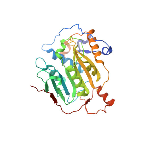
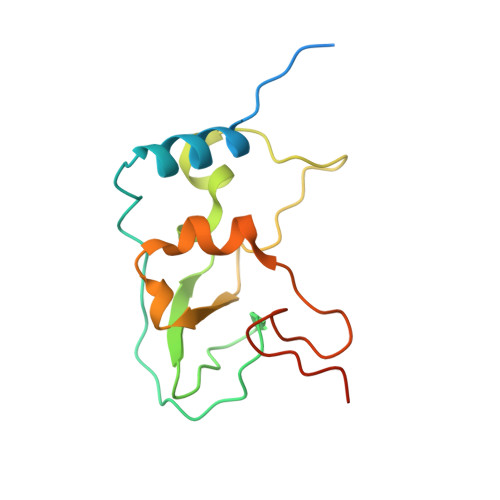
A metal ion orients SARS-CoV-2 mRNA to ensure accurate 2'-O methylation of its first nucleotide.

Viswanathan, T.Misra, A.Chan, S.H.Qi, S.Dai, N.Arya, S.Martinez-Sobrido, L.Gupta, Y.K.

(2021) Nat Commun 12: 3287-3287

  • DOI: https://doi.org/10.1038/s41467-021-23594-y
  • Primary Citation Related Structures: 
    7LW3, 7LW4

  • PubMed Abstract: 

    The SARS-CoV-2 nsp16/nsp10 enzyme complex modifies the 2'-OH of the first transcribed nucleotide of the viral mRNA by covalently attaching a methyl group to it. The 2'-O methylation of the first nucleotide converts the status of mRNA cap from Cap-0 to Cap-1, and thus, helps the virus evade immune surveillance in host cells. Here, we report two structures of nsp16/nsp10 representing pre- and post-release states of the RNA product (Cap-1). We observe overall widening of the enzyme upon product formation, and an inward twisting motion in the substrate binding region upon product release. These conformational changes reset the enzyme for the next round of catalysis. The structures also identify a unique binding mode and the importance of a divalent metal ion for 2'-O methylation. We also describe underlying structural basis for the perturbed enzymatic activity of a clinical variant of SARS-CoV-2, and a previous SARS-CoV outbreak strain.


  • Organizational Affiliation
    • Greehey Children's Cancer Research Institute, University of Texas Health at San Antonio, San Antonio, TX, USA.

Macromolecule Content 

  • Total Structure Weight: 50.09 kDa 
  • Atom Count: 3,444 
  • Modeled Residue Count: 413 
  • Deposited Residue Count: 437 
  • Unique protein chains: 2

Macromolecules

Find similar proteins by:|  3D Structure
Entity ID: 1
MoleculeChains  Sequence LengthOrganismDetailsImage
2'-O-methyltransferase298Severe acute respiratory syndrome coronavirus 2Mutation(s): 0 
Gene Names: rep1a-1b
EC: 2.1.1
UniProt
Find proteins for P0DTD1 (Severe acute respiratory syndrome coronavirus 2)
Explore P0DTD1 
Go to UniProtKB:  P0DTD1
Entity Groups
Sequence Clusters30% Identity50% Identity70% Identity90% Identity95% Identity100% Identity
UniProt GroupP0DTD1
Sequence Annotations
Expand
Reference Sequence
Find similar proteins by:|  3D Structure
Entity ID: 2
MoleculeChains  Sequence LengthOrganismDetailsImage
Non-structural protein 10139Severe acute respiratory syndrome coronavirus 2Mutation(s): 0 
Gene Names: rep1a-1b
UniProt
Find proteins for P0DTD1 (Severe acute respiratory syndrome coronavirus 2)
Explore P0DTD1 
Go to UniProtKB:  P0DTD1
Entity Groups
Sequence Clusters30% Identity50% Identity70% Identity90% Identity95% Identity100% Identity
UniProt GroupP0DTD1
Sequence Annotations
Expand
Reference Sequence

Small Molecules

Ligands 6 Unique
IDChains Name / Formula / InChI Key2D Diagram3D Interactions
YG4
(Subject of Investigation/LOI)

Query on YG4


Download:Ideal Coordinates CCD File
F [auth A][(2~{R},3~{R},4~{R},5~{R})-5-(6-azanyl-7,8-dihydropurin-9-yl)-2-[[[[[(2~{R},3~{S},4~{R},5~{R})-5-(2-azanyl-7-methyl-6-oxidanylidene-1,8-dihydropurin-9-yl)-3,4-bis(oxidanyl)oxolan-2-yl]methoxy-oxidanyl-phosphoryl]oxy-oxidanyl-phosphoryl]oxy-oxidanyl-phosphoryl]oxymethyl]-4-methoxy-oxolan-3-yl] [(2~{R},3~{S},4~{R},5~{S})-5-[2,4-bis(oxidanylidene)pyrimidin-1-yl]-3,4-bis(oxidanyl)oxolan-2-yl]methyl hydrogen phosphate
C31 H46 N12 O25 P4
POOGENYSODSFCS-SWDLWUBYSA-N
SAH
(Subject of Investigation/LOI)

Query on SAH


Download:Ideal Coordinates CCD File
E [auth A]S-ADENOSYL-L-HOMOCYSTEINE
C14 H20 N6 O5 S
ZJUKTBDSGOFHSH-WFMPWKQPSA-N
MES

Query on MES


Download:Ideal Coordinates CCD File
C [auth A]2-(N-MORPHOLINO)-ETHANESULFONIC ACID
C6 H13 N O4 S
SXGZJKUKBWWHRA-UHFFFAOYSA-N
ZN

Query on ZN


Download:Ideal Coordinates CCD File
H [auth B],
I [auth B]
ZINC ION
Zn
PTFCDOFLOPIGGS-UHFFFAOYSA-N
EDO

Query on EDO


Download:Ideal Coordinates CCD File
G [auth B]1,2-ETHANEDIOL
C2 H6 O2
LYCAIKOWRPUZTN-UHFFFAOYSA-N
MG
(Subject of Investigation/LOI)

Query on MG


Download:Ideal Coordinates CCD File
D [auth A]MAGNESIUM ION
Mg
JLVVSXFLKOJNIY-UHFFFAOYSA-N

Experimental Data & Validation

Experimental Data

  • Method: X-RAY DIFFRACTION
  • Resolution: 2.30 Å
  • R-Value Free:  0.251 (Depositor), 0.253 (DCC) 
  • R-Value Work:  0.222 (Depositor), 0.224 (DCC) 
  • R-Value Observed: 0.224 (Depositor) 
Space Group: P 31 2 1
Unit Cell:
Length ( Å )Angle ( ˚ )
a = 184.073α = 90
b = 184.073β = 90
c = 57.128γ = 120
Software Package:
Software NamePurpose
PHENIXrefinement
XDSdata reduction
Aimlessdata scaling
PHASERphasing

Structure Validation

View Full Validation Report


Ligand Structure Quality Assessment 


Entry History 

& Funding Information

Deposition Data


Funding OrganizationLocationGrant Number
Other privateUnited States--

Revision History  (Full details and data files)

  • Version 1.0: 2021-05-05
    Type: Initial release
  • Version 1.1: 2021-11-17
    Changes: Database references
  • Version 1.2: 2023-10-18
    Changes: Data collection, Refinement description