7LQ4

Rr (RsiG)2-(c-di-GMP)2-WhiG complex


Experimental Data Snapshot

  • Method: X-RAY DIFFRACTION
  • Resolution: 2.90 Å
  • R-Value Free: 0.268 
  • R-Value Work: 0.267 
  • R-Value Observed: 0.267 

wwPDB Validation   3D Report Full Report


Ligand Structure Quality Assessment 


This is version 1.2 of the entry. See complete history


Literature

Evolution of a sigma-(c-di-GMP)-anti-sigma switch.

Schumacher, M.A.Gallagher, K.A.Holmes, N.A.Chandra, G.Henderson, M.Kysela, D.T.Brennan, R.G.Buttner, M.J.

(2021) Proc Natl Acad Sci U S A 118

  • DOI: https://doi.org/10.1073/pnas.2105447118
  • Primary Citation of Related Structures:  
    7LQ2, 7LQ3, 7LQ4

  • PubMed Abstract: 

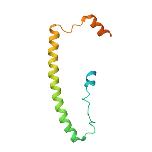
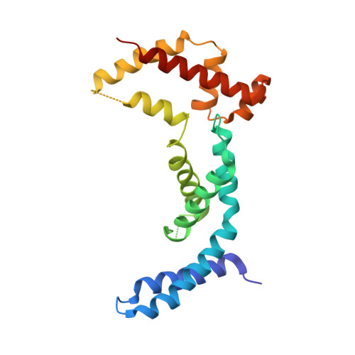
    Filamentous actinobacteria of the genus Streptomyces have a complex lifecycle involving the differentiation of reproductive aerial hyphae into spores. We recently showed c-di-GMP controls this transition by arming a unique anti-σ, RsiG, to bind the sporulation-specific σ, WhiG. The Streptomyces venezuelae RsiG-(c-di-GMP) 2 -WhiG structure revealed that a monomeric RsiG binds c-di-GMP via two E(X) 3 S(X) 2 R(X) 3 Q(X) 3 D repeat motifs, one on each helix of an antiparallel coiled-coil. Here we show that RsiG homologs are found scattered throughout the Actinobacteria. Strikingly, RsiGs from unicellular bacteria descending from the most basal branch of the Actinobacteria are small proteins containing only one c-di-GMP binding motif, yet still bind their WhiG partners. Our structure of a Rubrobacter radiotolerans (RsiG) 2 -(c-di-GMP) 2 -WhiG complex revealed that these single-motif RsiGs are able to form an antiparallel coiled-coil through homodimerization, thereby allowing them to bind c-di-GMP similar to the monomeric twin-motif RsiGs. Further data show that in the unicellular actinobacterium R. radiotolerans , the (RsiG) 2 -(c-di-GMP) 2 -WhiG regulatory switch controls type IV pilus expression. Phylogenetic analysis indicates the single-motif RsiGs likely represent the ancestral state and an internal gene-duplication event gave rise to the twin-motif RsiGs inherited elsewhere in the Actinobacteria. Thus, these studies show how the anti-σ RsiG has evolved through an intragenic duplication event from a small protein carrying a single c-di-GMP binding motif, which functions as a homodimer, to a larger protein carrying two c-di-GMP binding motifs, which functions as a monomer. Consistent with this, our structures reveal potential selective advantages of the monomeric twin-motif anti-σ factors.


  • Organizational Affiliation

    Department of Biochemistry, Duke University School of Medicine, Durham, NC 27710; maria.schumacher@duke.edu mark.buttner@jic.ac.uk.


Macromolecules
Find similar proteins by:  (by identity cutoff)  |  3D Structure
Entity ID: 1
MoleculeChains Sequence LengthOrganismDetailsImage
RsiGA [auth D],
B [auth T]
118Rubrobacter radiotoleransMutation(s): 0 
Gene Names: RradSPS_1442
UniProt
Find proteins for A0A023X3Z4 (Rubrobacter radiotolerans)
Explore A0A023X3Z4 
Go to UniProtKB:  A0A023X3Z4
Entity Groups  
Sequence Clusters30% Identity50% Identity70% Identity90% Identity95% Identity100% Identity
UniProt GroupA0A023X3Z4
Sequence Annotations
Expand
  • Reference Sequence
Find similar proteins by:  (by identity cutoff)  |  3D Structure
Entity ID: 2
MoleculeChains Sequence LengthOrganismDetailsImage
WhiGC [auth A]198Rubrobacter radiotoleransMutation(s): 0 
UniProt
Find proteins for A0A023X3C8 (Rubrobacter radiotolerans)
Explore A0A023X3C8 
Go to UniProtKB:  A0A023X3C8
Entity Groups  
Sequence Clusters30% Identity50% Identity70% Identity90% Identity95% Identity100% Identity
UniProt GroupA0A023X3C8
Sequence Annotations
Expand
  • Reference Sequence
Small Molecules
Ligands 1 Unique
IDChains Name / Formula / InChI Key2D Diagram3D Interactions
C2E
Query on C2E

Download Ideal Coordinates CCD File 
D,
E [auth T]
9,9'-[(2R,3R,3aS,5S,7aR,9R,10R,10aS,12S,14aR)-3,5,10,12-tetrahydroxy-5,12-dioxidooctahydro-2H,7H-difuro[3,2-d:3',2'-j][1,3,7,9,2,8]tetraoxadiphosphacyclododecine-2,9-diyl]bis(2-amino-1,9-dihydro-6H-purin-6-one)
C20 H24 N10 O14 P2
PKFDLKSEZWEFGL-MHARETSRSA-N
Experimental Data & Validation

Experimental Data

  • Method: X-RAY DIFFRACTION
  • Resolution: 2.90 Å
  • R-Value Free: 0.268 
  • R-Value Work: 0.267 
  • R-Value Observed: 0.267 
  • Space Group: P 21 21 21
Unit Cell:
Length ( Å )Angle ( ˚ )
a = 46.466α = 90
b = 81.621β = 90
c = 114.588γ = 90
Software Package:
Software NamePurpose
XDSdata reduction
SCALAdata scaling
MOLREPphasing
PHENIXrefinement
PDB_EXTRACTdata extraction

Structure Validation

View Full Validation Report


Ligand Structure Quality Assessment 


Entry History & Funding Information

Deposition Data


Funding OrganizationLocationGrant Number
National Institutes of Health/National Institute of General Medical Sciences (NIH/NIGMS)United StatesR35GM130290

Revision History  (Full details and data files)

  • Version 1.0: 2021-07-14
    Type: Initial release
  • Version 1.1: 2021-08-25
    Changes: Database references
  • Version 1.2: 2023-10-18
    Changes: Data collection, Refinement description