


SARS-CoV-2 Spike receptor-binding domain with a G485R mutation in complex with human ACE2
Weekley, C.M., Purcell, D.F.J., Parker, M.W.(2021) bioRxiv
Experimental Data Snapshot
Starting Model: experimental
View more details
(2021) bioRxiv
Entity ID: 1 | |||||
|---|---|---|---|---|---|
| Molecule | Chains | Sequence Length | Organism | Details | Image |
| Processed angiotensin-converting enzyme 2 | 596 | Homo sapiens | Mutation(s): 0 Gene Names: ACE2, UNQ868/PRO1885 EC: 3.4.17 (UniProt), 3.4.17.23 (UniProt) | 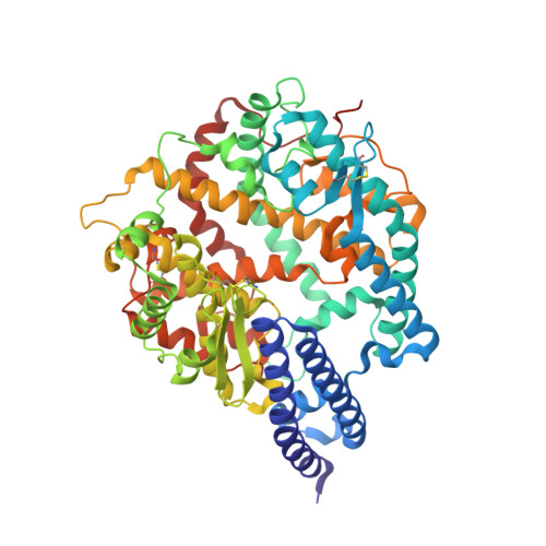 | |
UniProt & NIH Common Fund Data Resources | |||||
PHAROS: Q9BYF1 GTEx: ENSG00000130234 | |||||
Entity Groups | |||||
| Sequence Clusters | 30% Identity50% Identity70% Identity90% Identity95% Identity100% Identity | ||||
| UniProt Group | Q9BYF1 | ||||
Glycosylation | |||||
| Glycosylation Sites: 4 | Go to GlyGen: Q9BYF1-1 | ||||
Sequence AnnotationsExpand | |||||
Reference Sequence | |||||
Entity ID: 2 | |||||
|---|---|---|---|---|---|
| Molecule | Chains | Sequence Length | Organism | Details | Image |
| Spike protein S1 | 200 | Severe acute respiratory syndrome coronavirus 2 | Mutation(s): 1 Gene Names: S, 2 | 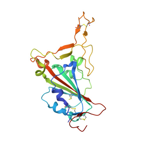 | |
UniProt | |||||
Entity Groups | |||||
| Sequence Clusters | 30% Identity50% Identity70% Identity90% Identity95% Identity100% Identity | ||||
| UniProt Group | P0DTC2 | ||||
Glycosylation | |||||
| Glycosylation Sites: 1 | Go to GlyGen: P0DTC2-1 | ||||
Sequence AnnotationsExpand | |||||
Reference Sequence | |||||
| Ligands 3 Unique | |||||
|---|---|---|---|---|---|
| ID | Chains | Name / Formula / InChI Key | 2D Diagram | 3D Interactions | |
| NAG Download:Ideal Coordinates CCD File | D [auth A], E [auth A], F [auth A], K [auth B] | 2-acetamido-2-deoxy-beta-D-glucopyranose C8 H15 N O6 OVRNDRQMDRJTHS-FMDGEEDCSA-N |  | ||
| ZN Download:Ideal Coordinates CCD File | J [auth A] | ZINC ION Zn PTFCDOFLOPIGGS-UHFFFAOYSA-N |  | ||
| EDO Download:Ideal Coordinates CCD File | G [auth A], H [auth A], I [auth A] | 1,2-ETHANEDIOL C2 H6 O2 LYCAIKOWRPUZTN-UHFFFAOYSA-N |  | ||
| Length ( Å ) | Angle ( ˚ ) |
|---|---|
| a = 65.606 | α = 90 |
| b = 112.176 | β = 90 |
| c = 145.989 | γ = 90 |
| Software Name | Purpose |
|---|---|
| PHENIX | refinement |
| XDS | data reduction |
| Aimless | data scaling |
| PHASER | phasing |
| PDB_EXTRACT | data extraction |


