7LJ1

Human Prx1-Srx Decameric Complex


Experimental Data Snapshot

  • Method: X-RAY DIFFRACTION
  • Resolution: 2.97 Å
  • R-Value Free: 0.230 
  • R-Value Work: 0.192 
  • R-Value Observed: 0.194 

wwPDB Validation   3D Report Full Report


Ligand Structure Quality Assessment 


This is version 1.2 of the entry. See complete history


Literature

Specificity of Human Sulfiredoxin for Reductant and Peroxiredoxin Oligomeric State.

Forshaw, T.E.Reisz, J.A.Nelson, K.J.Gumpena, R.Lawson, J.R.Jonsson, T.J.Wu, H.Clodfelter, J.E.Johnson, L.C.Furdui, C.M.Lowther, W.T.

(2021) Antioxidants (Basel) 10

  • DOI: https://doi.org/10.3390/antiox10060946
  • Primary Citation of Related Structures:  
    7LJ1

  • PubMed Abstract: 

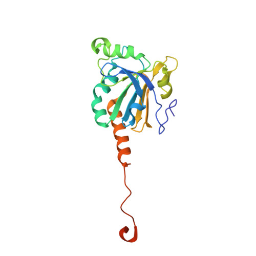
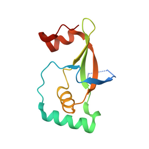
    Human peroxiredoxins (Prx) are a family of antioxidant enzymes involved in a myriad of cellular functions and diseases. During the reaction with peroxides (e.g., H 2 O 2 ), the typical 2-Cys Prxs change oligomeric structure between higher order (do)decamers and disulfide-linked dimers, with the hyperoxidized inactive state (-SO 2 H) favoring the multimeric structure of the reduced enzyme. Here, we present a study on the structural requirements for the repair of hyperoxidized 2-Cys Prxs by human sulfiredoxin (Srx) and the relative efficacy of physiological reductants hydrogen sulfide (H 2 S) and glutathione (GSH) in this reaction. The crystal structure of the toroidal Prx1-Srx complex shows an extended active site interface. The loss of this interface within engineered Prx2 and Prx3 dimers yielded variants more resistant to hyperoxidation and repair by Srx. Finally, we reveal for the first time Prx isoform-dependent use of and potential cooperation between GSH and H 2 S in supporting Srx activity.


  • Organizational Affiliation

    Department of Internal Medicine, Section on Molecular Medicine, Wake Forest School of Medicine, Medical Center Blvd., Winston-Salem, NC 27157, USA.


Macromolecules
Find similar proteins by:  (by identity cutoff)  |  3D Structure
Entity ID: 1
MoleculeChains Sequence LengthOrganismDetailsImage
Peroxiredoxin-1199Homo sapiensMutation(s): 3 
Gene Names: PRDX1PAGAPAGBTDPX2
EC: 1.11.1.24
UniProt & NIH Common Fund Data Resources
Find proteins for Q06830 (Homo sapiens)
Explore Q06830 
Go to UniProtKB:  Q06830
PHAROS:  Q06830
GTEx:  ENSG00000117450 
Entity Groups  
Sequence Clusters30% Identity50% Identity70% Identity90% Identity95% Identity100% Identity
UniProt GroupQ06830
Sequence Annotations
Expand
  • Reference Sequence
Find similar proteins by:  (by identity cutoff)  |  3D Structure
Entity ID: 2
MoleculeChains Sequence LengthOrganismDetailsImage
Sulfiredoxin-1109Homo sapiensMutation(s): 0 
Gene Names: SRXN1C20orf139SRXSRX1
EC: 1.8.98.2
UniProt & NIH Common Fund Data Resources
Find proteins for Q9BYN0 (Homo sapiens)
Explore Q9BYN0 
Go to UniProtKB:  Q9BYN0
PHAROS:  Q9BYN0
GTEx:  ENSG00000271303 
Entity Groups  
Sequence Clusters30% Identity50% Identity70% Identity90% Identity95% Identity100% Identity
UniProt GroupQ9BYN0
Sequence Annotations
Expand
  • Reference Sequence
Small Molecules
Ligands 5 Unique
IDChains Name / Formula / InChI Key2D Diagram3D Interactions
ATP (Subject of Investigation/LOI)
Query on ATP

Download Ideal Coordinates CCD File 
RB [auth c],
VB [auth g]
ADENOSINE-5'-TRIPHOSPHATE
C10 H16 N5 O13 P3
ZKHQWZAMYRWXGA-KQYNXXCUSA-N
PEG
Query on PEG

Download Ideal Coordinates CCD File 
CB [auth K]
DB [auth N]
EB [auth O]
IB [auth P]
KB [auth Q]
CB [auth K],
DB [auth N],
EB [auth O],
IB [auth P],
KB [auth Q],
NB [auth R],
SA [auth D],
WA [auth G]
DI(HYDROXYETHYL)ETHER
C4 H10 O3
MTHSVFCYNBDYFN-UHFFFAOYSA-N
PO4
Query on PO4

Download Ideal Coordinates CCD File 
AC [auth l]
BC [auth m]
CC [auth n]
DC [auth o]
EC [auth p]
AC [auth l],
BC [auth m],
CC [auth n],
DC [auth o],
EC [auth p],
FC [auth q],
GC [auth r],
HC [auth s],
IC [auth t],
PA [auth a],
QB [auth b],
SB [auth d],
TA [auth E],
TB [auth e],
UB [auth f],
WB [auth h],
XB [auth i],
YB [auth j],
ZB [auth k]
PHOSPHATE ION
O4 P
NBIIXXVUZAFLBC-UHFFFAOYSA-K
GOL
Query on GOL

Download Ideal Coordinates CCD File 
GB [auth O],
XA [auth H]
GLYCEROL
C3 H8 O3
PEDCQBHIVMGVHV-UHFFFAOYSA-N
EDO
Query on EDO

Download Ideal Coordinates CCD File 
AB [auth H]
BB [auth J]
FB [auth O]
HB [auth P]
JB [auth Q]
AB [auth H],
BB [auth J],
FB [auth O],
HB [auth P],
JB [auth Q],
LB [auth R],
MB [auth R],
OA [auth A],
OB [auth S],
PB [auth S],
QA [auth C],
RA [auth C],
UA [auth G],
VA [auth G],
YA [auth H],
ZA [auth H]
1,2-ETHANEDIOL
C2 H6 O2
LYCAIKOWRPUZTN-UHFFFAOYSA-N
Experimental Data & Validation

Experimental Data

  • Method: X-RAY DIFFRACTION
  • Resolution: 2.97 Å
  • R-Value Free: 0.230 
  • R-Value Work: 0.192 
  • R-Value Observed: 0.194 
  • Space Group: C 1 2 1
Unit Cell:
Length ( Å )Angle ( ˚ )
a = 330.834α = 90
b = 109.937β = 122.34
c = 260.143γ = 90
Software Package:
Software NamePurpose
REFMACrefinement
HKL-3000data reduction
HKL-3000data scaling
PHASERphasing

Structure Validation

View Full Validation Report


Ligand Structure Quality Assessment 


Entry History & Funding Information

Deposition Data


Funding OrganizationLocationGrant Number
National Institutes of Health/National Institute of General Medical Sciences (NIH/NIGMS)United StatesGM072866

Revision History  (Full details and data files)

  • Version 1.0: 2021-07-14
    Type: Initial release
  • Version 1.1: 2021-09-01
    Changes: Database references, Structure summary
  • Version 1.2: 2023-10-18
    Changes: Data collection, Refinement description