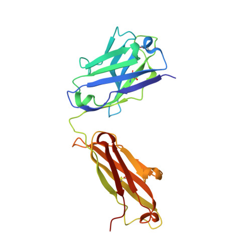
7LF7

Fab 6D12 bound to ApoL1 NTD


Experimental Data Snapshot

  • Method: X-RAY DIFFRACTION
  • Resolution: 2.03 Å
  • R-Value Free: 0.217 
  • R-Value Work: 0.189 
  • R-Value Observed: 0.190 

wwPDB Validation   3D Report Full Report


This is version 1.2 of the entry. See complete history


Literature

Structures of the ApoL1 and ApoL2 N-terminal domains reveal a non-classical four-helix bundle motif.

Ultsch, M.Holliday, M.J.Gerhardy, S.Moran, P.Scales, S.J.Gupta, N.Oltrabella, F.Chiu, C.Fairbrother, W.Eigenbrot, C.Kirchhofer, D.

(2021) Commun Biol 4: 916-916

  • DOI: https://doi.org/10.1038/s42003-021-02387-5
  • Primary Citation of Related Structures:  
    7L6K, 7LF7, 7LF8, 7LFA, 7LFB, 7LFD

  • PubMed Abstract: 

    Apolipoprotein L1 (ApoL1) is a circulating innate immunity protein protecting against trypanosome infection. However, two ApoL1 coding variants are associated with a highly increased risk of chronic kidney disease. Here we present X-ray and NMR structures of the N-terminal domain (NTD) of ApoL1 and of its closest relative ApoL2. In both proteins, four of the five NTD helices form a four-helix core structure which is different from the classical four-helix bundle and from the pore-forming domain of colicin A. The reactivity with a conformation-specific antibody and structural models predict that this four-helix motif is also present in the NTDs of ApoL3 and ApoL4, suggesting related functions within the small ApoL family. The long helix 5 of ApoL1 is conformationally flexible and contains the BH3-like region. This BH3-like α-helix resembles true BH3 domains only in sequence and structure but not in function, since it does not bind to the pro-survival members of the Bcl-2 family, suggesting a Bcl-2-independent role in cytotoxicity. These findings should expedite a more comprehensive structural and functional understanding of the ApoL immune protein family.


  • Organizational Affiliation

    Department of Structural Biology, Genentech Inc., South San Francisco, CA, USA.


Macromolecules
Find similar proteins by:  (by identity cutoff)  |  3D Structure
Entity ID: 1
MoleculeChains Sequence LengthOrganismDetailsImage
Fab 6D12 heavy chain225Homo sapiensMutation(s): 0 
Entity Groups  
Sequence Clusters30% Identity50% Identity70% Identity90% Identity95% Identity100% Identity
Sequence Annotations
Expand
  • Reference Sequence
Find similar proteins by:  (by identity cutoff)  |  3D Structure
Entity ID: 2
MoleculeChains Sequence LengthOrganismDetailsImage
Fab 6D12 light chainB,
E [auth L]
214Homo sapiensMutation(s): 0 
Entity Groups  
Sequence Clusters30% Identity50% Identity70% Identity90% Identity95% Identity100% Identity
Sequence Annotations
Expand
  • Reference Sequence
Find similar proteins by:  (by identity cutoff)  |  3D Structure
Entity ID: 3
MoleculeChains Sequence LengthOrganismDetailsImage
Fab 6D12 heavy chainC [auth H]225Homo sapiensMutation(s): 0 
Entity Groups  
Sequence Clusters30% Identity50% Identity70% Identity90% Identity95% Identity100% Identity
Sequence Annotations
Expand
  • Reference Sequence
Find similar proteins by:  (by identity cutoff)  |  3D Structure
Entity ID: 4
MoleculeChains Sequence LengthOrganismDetailsImage
Apolipoprotein L1D [auth K],
F [auth M]
130Homo sapiensMutation(s): 0 
Gene Names: APOL1APOL
Membrane Entity: Yes 
UniProt & NIH Common Fund Data Resources
Find proteins for O14791 (Homo sapiens)
Explore O14791 
Go to UniProtKB:  O14791
PHAROS:  O14791
GTEx:  ENSG00000100342 
Entity Groups  
Sequence Clusters30% Identity50% Identity70% Identity90% Identity95% Identity100% Identity
UniProt GroupO14791
Sequence Annotations
Expand
  • Reference Sequence
Experimental Data & Validation

Experimental Data

  • Method: X-RAY DIFFRACTION
  • Resolution: 2.03 Å
  • R-Value Free: 0.217 
  • R-Value Work: 0.189 
  • R-Value Observed: 0.190 
  • Space Group: P 43
Unit Cell:
Length ( Å )Angle ( ˚ )
a = 131.103α = 90
b = 131.103β = 90
c = 87.164γ = 90
Software Package:
Software NamePurpose
PHENIXrefinement
PDB_EXTRACTdata extraction
XDSdata reduction
Aimlessdata scaling
PHASERphasing

Structure Validation

View Full Validation Report


Entry History 

Deposition Data

Revision History  (Full details and data files)

  • Version 1.0: 2021-08-04
    Type: Initial release
  • Version 1.1: 2022-02-16
    Changes: Database references
  • Version 1.2: 2023-10-18
    Changes: Data collection, Refinement description