7L6L

Crystal Structure of the DNA-binding Transcriptional Repressor DeoR from Escherichia coli str. K-12


Experimental Data Snapshot

  • Method: X-RAY DIFFRACTION
  • Resolution: 1.75 Å
  • R-Value Free: 0.183 
  • R-Value Work: 0.158 
  • R-Value Observed: 0.159 

wwPDB Validation   3D Report Full Report


This is version 1.0 of the entry. See complete history


Literature

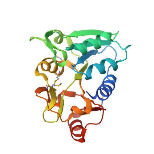
Crystal Structure of the DNA-binding Transcriptional Repressor DeoR from Escherichia coli str. K-12.

Minasov, G.Shuvalova, L.Kiryukhina, O.Dubrovska, I.Wiersum, G.Satchell, K.J.F.Center for Structural Genomics of Infectious Diseases (CSGID)

To be published.

Macromolecules
Find similar proteins by:  (by identity cutoff)  |  3D Structure
Entity ID: 1
MoleculeChains Sequence LengthOrganismDetailsImage
Deoxyribose operon repressor
A, B, C, D, E
A, B, C, D, E, F
186Escherichia coli K-12Mutation(s): 0 
Gene Names: deoRnucRb0840JW0824
UniProt
Find proteins for P0ACK5 (Escherichia coli (strain K12))
Explore P0ACK5 
Go to UniProtKB:  P0ACK5
Entity Groups  
Sequence Clusters30% Identity50% Identity70% Identity90% Identity95% Identity100% Identity
UniProt GroupP0ACK5
Sequence Annotations
Expand
  • Reference Sequence
Small Molecules
Ligands 3 Unique
IDChains Name / Formula / InChI Key2D Diagram3D Interactions
SO4
Query on SO4

Download Ideal Coordinates CCD File 
AA [auth D]
CA [auth E]
DA [auth E]
FA [auth F]
GA [auth F]
AA [auth D],
CA [auth E],
DA [auth E],
FA [auth F],
GA [auth F],
H [auth A],
HA [auth F],
I [auth A],
J [auth A],
M [auth B],
N [auth B],
O [auth B],
P [auth B],
Q [auth B],
T [auth C],
U [auth C],
V [auth C],
X [auth D],
Y [auth D],
Z [auth D]
SULFATE ION
O4 S
QAOWNCQODCNURD-UHFFFAOYSA-L
EDO
Query on EDO

Download Ideal Coordinates CCD File 
EA [auth F],
G [auth A],
L [auth B],
S [auth C]
1,2-ETHANEDIOL
C2 H6 O2
LYCAIKOWRPUZTN-UHFFFAOYSA-N
CL
Query on CL

Download Ideal Coordinates CCD File 
BA [auth E],
K [auth B],
R [auth C],
W [auth D]
CHLORIDE ION
Cl
VEXZGXHMUGYJMC-UHFFFAOYSA-M
Modified Residues  1 Unique
IDChains TypeFormula2D DiagramParent
MSE
Query on MSE
A, B, C, D, E
A, B, C, D, E, F
L-PEPTIDE LINKINGC5 H11 N O2 SeMET
Experimental Data & Validation

Experimental Data

  • Method: X-RAY DIFFRACTION
  • Resolution: 1.75 Å
  • R-Value Free: 0.183 
  • R-Value Work: 0.158 
  • R-Value Observed: 0.159 
  • Space Group: P 1 21 1
Unit Cell:
Length ( Å )Angle ( ˚ )
a = 70.751α = 90
b = 64.734β = 97.94
c = 140.71γ = 90
Software Package:
Software NamePurpose
REFMACrefinement
PDB_EXTRACTdata extraction
HKL-3000data reduction
HKL-3000data scaling
HKL-3000phasing

Structure Validation

View Full Validation Report


Entry History 

Revision History  (Full details and data files)

  • Version 1.0: 2021-12-01
    Type: Initial release