


Crystal Structure of Cytidylate kinase from Encephalitozoon cuniculi GB-M1 in complex with two CDP molecules
Abendroth, J.A., Fox III, D., Lorimer, D.D., Horanyi, P.S., Edwards, T.E.To be published.
Experimental Data Snapshot
Entity ID: 1 | |||||
|---|---|---|---|---|---|
| Molecule | Chains | Sequence Length | Organism | Details | Image |
| Cytidylate kinase | 229 | Encephalitozoon cuniculi GB-M1 | Mutation(s): 0 Gene Names: ECU03_1270 EC: 2.7.4.25 | 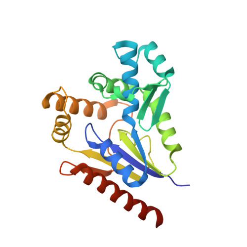 | |
UniProt | |||||
Entity Groups | |||||
| Sequence Clusters | 30% Identity50% Identity70% Identity90% Identity95% Identity100% Identity | ||||
| UniProt Group | Q8SS83 | ||||
Sequence AnnotationsExpand | |||||
Reference Sequence | |||||
| Ligands 3 Unique | |||||
|---|---|---|---|---|---|
| ID | Chains | Name / Formula / InChI Key | 2D Diagram | 3D Interactions | |
| CDP (Subject of Investigation/LOI) Download:Ideal Coordinates CCD File | B [auth A], C [auth A] | CYTIDINE-5'-DIPHOSPHATE C9 H15 N3 O11 P2 ZWIADYZPOWUWEW-XVFCMESISA-N |  | ||
| SO4 Download:Ideal Coordinates CCD File | D [auth A] | SULFATE ION O4 S QAOWNCQODCNURD-UHFFFAOYSA-L |  | ||
| EDO Download:Ideal Coordinates CCD File | E [auth A] | 1,2-ETHANEDIOL C2 H6 O2 LYCAIKOWRPUZTN-UHFFFAOYSA-N |  | ||
| Length ( Å ) | Angle ( ˚ ) |
|---|---|
| a = 48.52 | α = 90 |
| b = 46.87 | β = 112.592 |
| c = 53.83 | γ = 90 |
| Software Name | Purpose |
|---|---|
| XDS | data reduction |
| XSCALE | data scaling |
| PHENIX | refinement |
| PDB_EXTRACT | data extraction |
| PHASER | phasing |
| PARROT | phasing |
| ARP/wARP | model building |
| Coot | model building |


