7L35

Human DNA Ligase 1 - R771W nicked DNA complex


Experimental Data Snapshot

  • Method: X-RAY DIFFRACTION
  • Resolution: 2.00 Å
  • R-Value Free: 0.195 
  • R-Value Work: 0.163 
  • R-Value Observed: 0.164 

wwPDB Validation   3D Report Full Report


Ligand Structure Quality Assessment 


This is version 1.3 of the entry. See complete history


Literature

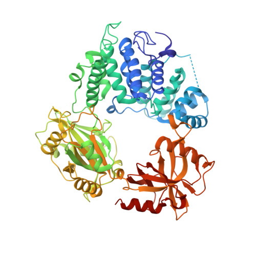
LIG1 syndrome mutations remodel a cooperative network of ligand binding interactions to compromise ligation efficiency.

Jurkiw, T.J.Tumbale, P.P.Schellenberg, M.J.Cunningham-Rundles, C.Williams, R.S.O'Brien, P.J.

(2021) Nucleic Acids Res 49: 1619-1630

  • DOI: https://doi.org/10.1093/nar/gkaa1297
  • Primary Citation of Related Structures:  
    7L34, 7L35

  • PubMed Abstract: 

    Human DNA ligase I (LIG1) is the main replicative ligase and it also seals DNA breaks to complete DNA repair and recombination pathways. Immune compromised patients harbor hypomorphic LIG1 alleles encoding substitutions of conserved arginine residues, R771W and R641L, that compromise LIG1 activity through poorly defined mechanisms. To understand the molecular basis of LIG1 syndrome mutations, we determined high resolution X-ray structures and performed systematic biochemical characterization of LIG1 mutants using steady-state and pre-steady state kinetic approaches. Our results unveil a cooperative network of plastic DNA-LIG1 interactions that connect DNA substrate engagement with productive binding of Mg2+ cofactors for catalysis. LIG1 syndrome mutations destabilize this network, compromising Mg2+ binding affinity, decreasing ligation efficiency, and leading to elevated abortive ligation that may underlie the disease pathology. These findings provide novel insights into the fundamental mechanism by which DNA ligases engage with a nicked DNA substrate, and they suggest that disease pathology of LIG1 syndrome could be modulated by Mg2+ levels.


  • Organizational Affiliation

    Department of Biological Chemistry, University of Michigan School of Medicine, Ann Arbor, MI, 48109, USA.


Macromolecules

Find similar proteins by:  (by identity cutoff)  |  3D Structure
Entity ID: 1
MoleculeChains Sequence LengthOrganismDetailsImage
DNA ligase 1647Homo sapiensMutation(s): 1 
Gene Names: LIG1
EC: 6.5.1.1
UniProt & NIH Common Fund Data Resources
Find proteins for P18858 (Homo sapiens)
Explore P18858 
Go to UniProtKB:  P18858
PHAROS:  P18858
GTEx:  ENSG00000105486 
Entity Groups  
Sequence Clusters30% Identity50% Identity70% Identity90% Identity95% Identity100% Identity
UniProt GroupP18858
Sequence Annotations
Expand
  • Reference Sequence

Find similar nucleic acids by:  Sequence   |   3D Structure  

Entity ID: 2
MoleculeChains LengthOrganismImage
DNA (5'-D(*GP*CP*TP*GP*AP*TP*GP*CP*GP*TP*C)-3')11Homo sapiens
Sequence Annotations
Expand
  • Reference Sequence

Find similar nucleic acids by:  Sequence   |   3D Structure  

Entity ID: 3
MoleculeChains LengthOrganismImage
DNA (5'-D(P*GP*TP*CP*GP*GP*AP*C)-3')7Homo sapiens
Sequence Annotations
Expand
  • Reference Sequence

Find similar nucleic acids by:  Sequence   |   3D Structure  

Entity ID: 4
MoleculeChains LengthOrganismImage
DNA (5'-D(*GP*TP*CP*CP*GP*AP*CP*GP*AP*CP*GP*CP*AP*TP*CP*AP*GP*C)-3')18Homo sapiens
Sequence Annotations
Expand
  • Reference Sequence
Experimental Data & Validation

Experimental Data

  • Method: X-RAY DIFFRACTION
  • Resolution: 2.00 Å
  • R-Value Free: 0.195 
  • R-Value Work: 0.163 
  • R-Value Observed: 0.164 
  • Space Group: P 21 21 21
Unit Cell:
Length ( Å )Angle ( ˚ )
a = 71.33α = 90
b = 100.523β = 90
c = 116.088γ = 90
Software Package:
Software NamePurpose
PHENIXrefinement
PDB_EXTRACTdata extraction
HKL-2000data reduction
HKL-2000data scaling
PHASERphasing

Structure Validation

View Full Validation Report


Ligand Structure Quality Assessment 


Entry History & Funding Information

Deposition Data


Funding OrganizationLocationGrant Number
National Institutes of Health/National Institute of Environmental Health Sciences (NIH/NIEHS)United StatesZ01ES102765

Revision History  (Full details and data files)

  • Version 1.0: 2021-01-13
    Type: Initial release
  • Version 1.1: 2021-01-27
    Changes: Database references
  • Version 1.2: 2021-03-03
    Changes: Database references
  • Version 1.3: 2023-10-18
    Changes: Data collection, Database references, Refinement description