7KYW | pdb_00007kyw

Crystal structure of timothy grass allergen Phl p 12.0101 reveals an unusual profilin dimer


Experimental Data Snapshot

  • Method: X-RAY DIFFRACTION
  • Resolution: 2.30 Å
  • R-Value Free: 
    0.241 (Depositor), 0.253 (DCC) 
  • R-Value Work: 
    0.197 (Depositor) 
  • R-Value Observed: 
    0.200 (Depositor) 

Starting Model: experimental
View more details

wwPDB Validation 3D Report Full Report

Validation slider image for 7KYW

This is version 1.2 of the entry. See complete history

Literature

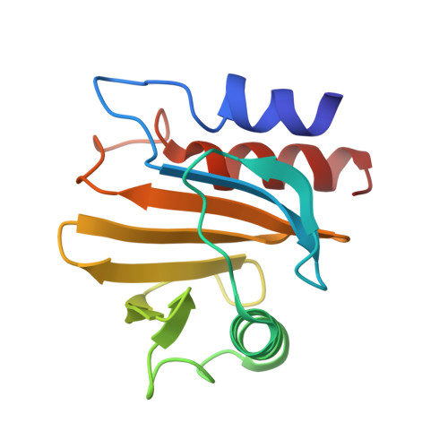
Crystal structure of timothy grass allergen Phl p 12.0101 reveals an unusual profilin dimer.

O'Malley, A.Kapingidza, A.B.Hyduke, N.Dolamore, C.Kowal, K.Chruszcz, M.

(2021) Acta Biochim Pol 68: 15-22

  • DOI: https://doi.org/10.18388/abp.2020_5587
  • Primary Citation Related Structures: 
    7KYW

  • PubMed Abstract: 

    Timothy grass pollen is a source of potent allergens. Among them, Phl p 1 and Phl p 5 are thought to be the most important, as a majority of timothy grass-allergic individuals have IgE antibodies directed against these two allergens. The profilin from timothy grass (Phl p 12) has been registered as a minor allergen, with up to 35% of individuals in populations of grass pollen allergic patients showing IgE binding to Phl p 12. Profilins are primarily minor allergens and are known for a high likelihood of co-sensitization as well as cross-reactivity situations caused by their sequence and structure similarity. The crystal structure of Phl p 12.0101 was determined and it revealed that this allergen may form an unusual dimer not previously observed among any profilins. For example, the Phl p 12 dimer has a completely different geometry and interface when compared with the latex profilin (Hev b 8) dimer that has its crystal structure determined. The structure of Phl p 12.0101 is described in the context of allergenic sensitization and allergy diagnostics. Moreover, the structure of the Phl p 12.0101 dimer is discussed, taking into account the production of recombinant allergens and their storage.


  • Organizational Affiliation
    • Department of Chemistry and Biochemistry, University of South Carolina, Columbia, SC 29208, USA.

Macromolecule Content 

  • Total Structure Weight: 14.37 kDa 
  • Atom Count: 1,029 
  • Modeled Residue Count: 131 
  • Deposited Residue Count: 131 
  • Unique protein chains: 1

Macromolecules

Find similar proteins by:|  3D Structure
Entity ID: 1
MoleculeChains  Sequence LengthOrganismDetailsImage
Profilin-1131Phleum pratenseMutation(s): 0 
Gene Names: PRO1PHLPXI
UniProt
Find proteins for P35079 (Phleum pratense)
Explore P35079 
Go to UniProtKB:  P35079
Entity Groups
Sequence Clusters30% Identity50% Identity70% Identity90% Identity95% Identity100% Identity
UniProt GroupP35079
Sequence Annotations
Expand
Reference Sequence

Small Molecules

Ligands 1 Unique
IDChains Name / Formula / InChI Key2D Diagram3D Interactions
CIT

Query on CIT


Download:Ideal Coordinates CCD File
B [auth A]CITRIC ACID
C6 H8 O7
KRKNYBCHXYNGOX-UHFFFAOYSA-N

Experimental Data & Validation

Experimental Data

  • Method: X-RAY DIFFRACTION
  • Resolution: 2.30 Å
  • R-Value Free:  0.241 (Depositor), 0.253 (DCC) 
  • R-Value Work:  0.197 (Depositor) 
  • R-Value Observed: 0.200 (Depositor) 
Space Group: P 31 2 1
Unit Cell:
Length ( Å )Angle ( ˚ )
a = 56.344α = 90
b = 56.344β = 90
c = 94.379γ = 120
Software Package:
Software NamePurpose
REFMACrefinement
PDB_EXTRACTdata extraction
HKL-3000data reduction
HKL-3000data scaling
MOLREPphasing

Structure Validation

View Full Validation Report


Entry History 

& Funding Information

Deposition Data


Funding OrganizationLocationGrant Number
National Institutes of Health/National Institute Of Allergy and Infectious Diseases (NIH/NIAID)United States2R01AI077653-10A1

Revision History  (Full details and data files)

  • Version 1.0: 2021-03-31
    Type: Initial release
  • Version 1.1: 2023-10-18
    Changes: Data collection, Database references, Refinement description
  • Version 1.2: 2024-11-06
    Changes: Structure summary