7KVV

Crystal structure of Squash RNA aptamer in complex with DFHBI-1T


Experimental Data Snapshot

  • Method: X-RAY DIFFRACTION
  • Resolution: 2.85 Å
  • R-Value Free: 0.245 
  • R-Value Work: 0.221 
  • R-Value Observed: 0.223 

wwPDB Validation   3D Report Full Report


Ligand Structure Quality Assessment 


This is version 1.2 of the entry. See complete history


Literature

The fluorescent aptamer Squash extensively repurposes the adenine riboswitch fold.

Truong, L.Kooshapur, H.Dey, S.K.Li, X.Tjandra, N.Jaffrey, S.R.Ferre-D'Amare, A.R.

(2022) Nat Chem Biol 18: 191-198

  • DOI: https://doi.org/10.1038/s41589-021-00931-2
  • Primary Citation of Related Structures:  
    7KVT, 7KVU, 7KVV

  • PubMed Abstract: 

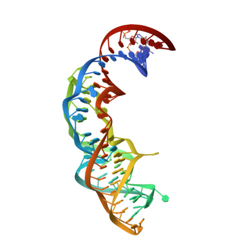
    Squash is an RNA aptamer that strongly activates the fluorescence of small-molecule analogs of the fluorophore of green fluorescent protein (GFP). Unlike other fluorogenic aptamers, isolated de novo from random-sequence RNA, Squash was evolved from the bacterial adenine riboswitch to leverage its optimized in vivo folding and stability. We now report the 2.7-Å resolution cocrystal structure of fluorophore-bound Squash, revealing that while the overall fold of the riboswitch is preserved, the architecture of the ligand-binding core is dramatically transformed. Unlike previously characterized aptamers that activate GFP-derived fluorophores, Squash does not harbor a G-quadruplex, sandwiching its fluorophore between a base triple and a noncanonical base quadruple in a largely apolar pocket. The expanded structural core of Squash allows it to recognize unnatural fluorophores that are larger than the simple purine ligand of the parental adenine riboswitch, and suggests that stable RNA scaffolds can tolerate larger variation than has hitherto been appreciated.


  • Organizational Affiliation

    Biochemistry and Biophysics Center, National Heart, Lung, and Blood Institute, Bethesda, MD, USA.


Macromolecules
Find similar nucleic acids by:  (by identity cutoff)  |  3D Structure
Entity ID: 1
MoleculeChains LengthOrganismImage
Squash RNA aptamer bound to DFHOA [auth D]83synthetic construct
Sequence Annotations
Expand
  • Reference Sequence
Small Molecules
Ligands 2 Unique
IDChains Name / Formula / InChI Key2D Diagram3D Interactions
747
Query on 747

Download Ideal Coordinates CCD File 
C [auth D](5Z)-5-[(3,5-difluoro-4-hydroxyphenyl)methylidene]-2-[(E)-(hydroxyimino)methyl]-3-methyl-3,5-dihydro-4H-imidazol-4-one
C12 H9 F2 N3 O3
MZFHSQVSFUWKDU-TUUOAFJKSA-N
MG
Query on MG

Download Ideal Coordinates CCD File 
B [auth D],
D
MAGNESIUM ION
Mg
JLVVSXFLKOJNIY-UHFFFAOYSA-N
Experimental Data & Validation

Experimental Data

  • Method: X-RAY DIFFRACTION
  • Resolution: 2.85 Å
  • R-Value Free: 0.245 
  • R-Value Work: 0.221 
  • R-Value Observed: 0.223 
  • Space Group: P 42 21 2
Unit Cell:
Length ( Å )Angle ( ˚ )
a = 104.271α = 90
b = 104.271β = 90
c = 52.107γ = 90
Software Package:
Software NamePurpose
PHENIXrefinement

Structure Validation

View Full Validation Report


Ligand Structure Quality Assessment 


Entry History & Funding Information

Deposition Data


Funding OrganizationLocationGrant Number
National Institutes of Health/National Heart, Lung, and Blood Institute (NIH/NHLBI)United States--

Revision History  (Full details and data files)

  • Version 1.0: 2022-01-19
    Type: Initial release
  • Version 1.1: 2022-02-16
    Changes: Database references
  • Version 1.2: 2023-10-18
    Changes: Data collection, Refinement description