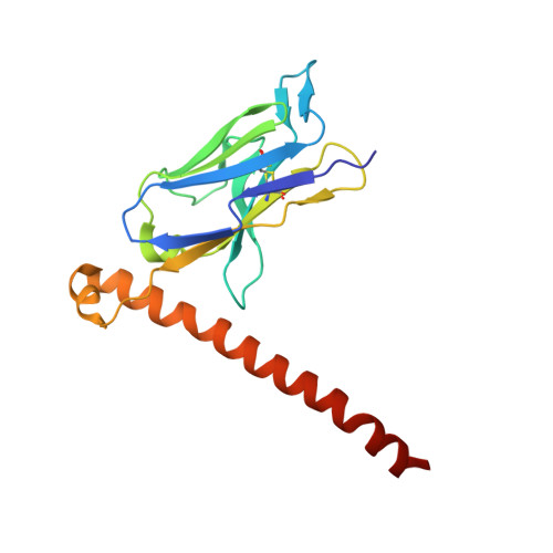
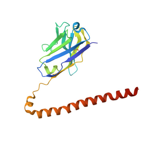
7KM6 | pdb_00007km6

APOBEC3B antibody 5G7 Fv-clasp

  • Classification: IMMUNE SYSTEM
  • Organism(s): Homo sapiens
  • Expression System: Escherichia coli
  • Mutation(s): No 

  • Deposited: 2020-11-02 Released: 2021-05-12 
  • Deposition Author(s): Tang, H., Shi, K., Aihara, H.
  • Funding Organization(s): National Institutes of Health/National Institute of General Medical Sciences (NIH/NIGMS), National Natural Science Foundation of China (NSFC)

Experimental Data Snapshot

  • Method: X-RAY DIFFRACTION
  • Resolution: 1.67 Å
  • R-Value Free: 
    0.238 (Depositor), 0.231 (DCC) 
  • R-Value Work: 
    0.189 (Depositor), 0.184 (DCC) 
  • R-Value Observed: 
    0.191 (Depositor) 

Starting Model: experimental
View more details

wwPDB Validation 3D Report Full Report

Validation slider image for 7KM6

This is version 1.2 of the entry. See complete history. 

Literature

Structural Characterization of a Minimal Antibody against Human APOBEC3B.

Tang, H., Demir, O., Kurniawan, F., Brown, W.L., Shi, K., Moeller, N.H., Carpenter, M.A., Belica, C., Orellana, K., Du, G., LeBeau, A.M., Amaro, R.E., Harris, R.S., Aihara, H.

(2021) Viruses 13

  • DOI: https://doi.org/10.3390/v13040663
  • Primary Citation Related Structures: 
    7KM6

  • PubMed Abstract: 

    APOBEC3B (A3B) is one of seven human APOBEC3 DNA cytosine deaminases that restrict viral infections as part of the overall innate immune response, but it also plays a major role in tumor evolution by mutating genomic DNA. Given the importance of A3B as a restriction factor of viral infections and as a driver of multiple human cancers, selective antibodies against A3B are highly desirable for its specific detection in various research and possibly diagnostic applications. Here, we describe a high-affinity minimal antibody, designated 5G7, obtained via a phage display screening against the C-terminal catalytic domain (ctd) of A3B. 5G7 also binds APOBEC3A that is highly homologous to A3Bctd but does not bind the catalytic domain of APOBEC3G, another Z1-type deaminase domain. The crystal structure of 5G7 shows a canonical arrangement of the heavy and light chain variable domains, with their complementarity-determining region (CDR) loops lining an antigen-binding cleft that accommodates a pair of α-helices. To understand the mechanism of A3Bctd recognition by 5G7, we used the crystal structures of A3Bctd and 5G7 as templates and computationally predicted the A3B-5G7 complex structure. Stable binding poses obtained by the simulation were further tested by site-directed mutagenesis and in vitro binding analyses. These studies mapped the epitope for 5G7 to a portion of C-terminal α6 helix of A3Bctd, with Arg374 playing an essential role. The same region of A3Bctd was used previously as a peptide antigen for generating a rabbit monoclonal antibody (mAb 5210-87-13), suggesting that this region is particularly immunogenic and that these antibodies from very different origins may share similar binding modes. Our studies provide a platform for the development of selective antibodies against A3B and other APOBEC3 family enzymes.


  • Organizational Affiliation: 
    • Department of Biochemistry, Molecular Biology, and Biophysics, University of Minnesota, Minneapolis, MN 55455, USA.

Macromolecule Content 

  • Total Structure Weight: 78.31 kDa 
  • Atom Count: 5,951 
  • Modeled Residue Count: 672 
  • Deposited Residue Count: 684 
  • Unique protein chains: 2

Macromolecules

Find similar proteins by:|  3D Structure
Entity ID: 1
MoleculeChains  Sequence LengthOrganismDetailsImage
5G7 human monoclonal FAB light chainA [auth L],
C [auth l]
166Homo sapiensMutation(s): 0 
Entity Groups
Sequence Clusters30% Identity50% Identity70% Identity90% Identity95% Identity100% Identity
Sequence Annotations
Expand
Reference Sequence
Find similar proteins by:|  3D Structure
Entity ID: 2
MoleculeChains  Sequence LengthOrganismDetailsImage
5G7 human monoclonal FAB heavy chainB [auth H],
D [auth h]
176Homo sapiensMutation(s): 0 
Entity Groups
Sequence Clusters30% Identity50% Identity70% Identity90% Identity95% Identity100% Identity
Sequence Annotations
Expand
Reference Sequence

Small Molecules

Ligands 3 Unique
IDChains Name / Formula / InChI Key2D Diagram3D Interactions
EDO

Query on EDO


Download:Ideal Coordinates CCD File
E [auth L]
F [auth L]
H
J [auth H]
K [auth H]
E [auth L],
F [auth L],
H,
J [auth H],
K [auth H],
L [auth H],
P [auth h],
Q [auth h],
R [auth h],
S [auth h]
1,2-ETHANEDIOL
C2 H6 O2
LYCAIKOWRPUZTN-UHFFFAOYSA-N
CL

Query on CL


Download:Ideal Coordinates CCD File
G [auth L],
M [auth l],
N [auth l]
CHLORIDE ION
Cl
VEXZGXHMUGYJMC-UHFFFAOYSA-M
NA

Query on NA


Download:Ideal Coordinates CCD File
I [auth H],
O [auth l]
SODIUM ION
Na
FKNQFGJONOIPTF-UHFFFAOYSA-N

Experimental Data & Validation

Experimental Data

  • Method: X-RAY DIFFRACTION
  • Resolution: 1.67 Å
  • R-Value Free:  0.238 (Depositor), 0.231 (DCC) 
  • R-Value Work:  0.189 (Depositor), 0.184 (DCC) 
  • R-Value Observed: 0.191 (Depositor) 
Space Group: P 1 21 1
Unit Cell:
Length ( Å )Angle ( ˚ )
a = 54.074α = 90
b = 68.823β = 107.05
c = 88.759γ = 90
Software Package:
Software NamePurpose
PHENIXrefinement
Aimlessdata scaling
PDB_EXTRACTdata extraction
XDSdata reduction
PHASERphasing

Structure Validation

View Full Validation Report


Entry History 

& Funding Information

Deposition Data


Funding OrganizationLocationGrant Number
National Institutes of Health/National Institute of General Medical Sciences (NIH/NIGMS)United StatesGM118047
National Natural Science Foundation of China (NSFC)China31470160

Revision History  (Full details and data files)

  • Version 1.0: 2021-05-12
    Type: Initial release
  • Version 1.1: 2023-10-18
    Changes: Data collection, Database references, Refinement description
  • Version 1.2: 2024-10-09
    Changes: Structure summary