Structural Characterization of a Minimal Antibody against Human APOBEC3B.
Tang, H., Demir, O., Kurniawan, F., Brown, W.L., Shi, K., Moeller, N.H., Carpenter, M.A., Belica, C., Orellana, K., Du, G., LeBeau, A.M., Amaro, R.E., Harris, R.S., Aihara, H.(2021) Viruses 13
- PubMed: 33921405 Search on PubMedSearch on PubMed Central
- DOI: https://doi.org/10.3390/v13040663
- Primary Citation Related Structures:
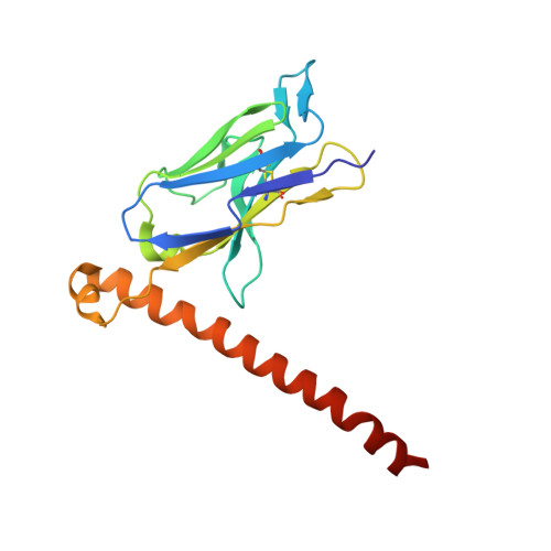
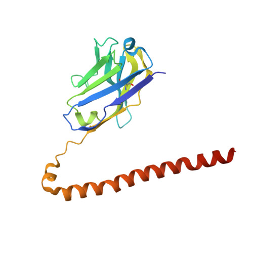
7KM6 - PubMed Abstract:
APOBEC3B (A3B) is one of seven human APOBEC3 DNA cytosine deaminases that restrict viral infections as part of the overall innate immune response, but it also plays a major role in tumor evolution by mutating genomic DNA. Given the importance of A3B as a restriction factor of viral infections and as a driver of multiple human cancers, selective antibodies against A3B are highly desirable for its specific detection in various research and possibly diagnostic applications. Here, we describe a high-affinity minimal antibody, designated 5G7, obtained via a phage display screening against the C-terminal catalytic domain (ctd) of A3B. 5G7 also binds APOBEC3A that is highly homologous to A3Bctd but does not bind the catalytic domain of APOBEC3G, another Z1-type deaminase domain. The crystal structure of 5G7 shows a canonical arrangement of the heavy and light chain variable domains, with their complementarity-determining region (CDR) loops lining an antigen-binding cleft that accommodates a pair of α-helices. To understand the mechanism of A3Bctd recognition by 5G7, we used the crystal structures of A3Bctd and 5G7 as templates and computationally predicted the A3B-5G7 complex structure. Stable binding poses obtained by the simulation were further tested by site-directed mutagenesis and in vitro binding analyses. These studies mapped the epitope for 5G7 to a portion of C-terminal α6 helix of A3Bctd, with Arg374 playing an essential role. The same region of A3Bctd was used previously as a peptide antigen for generating a rabbit monoclonal antibody (mAb 5210-87-13), suggesting that this region is particularly immunogenic and that these antibodies from very different origins may share similar binding modes. Our studies provide a platform for the development of selective antibodies against A3B and other APOBEC3 family enzymes.
- Department of Biochemistry, Molecular Biology, and Biophysics, University of Minnesota, Minneapolis, MN 55455, USA.
Organizational Affiliation: