7KBC

The crystal structure of the 2009/H1N1/California PA endonuclease mutant E119D (construct with truncated loop 51-72) in complex with baloxavir acid


Experimental Data Snapshot

  • Method: X-RAY DIFFRACTION
  • Resolution: 2.25 Å
  • R-Value Free: 0.228 
  • R-Value Work: 0.195 
  • R-Value Observed: 0.197 

wwPDB Validation   3D Report Full Report


Ligand Structure Quality Assessment 


This is version 1.2 of the entry. See complete history


Literature

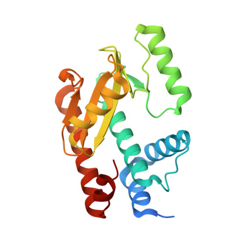
Structural insights into the substrate specificity of the endonuclease activity of the influenza virus cap-snatching mechanism.

Kumar, G.Cuypers, M.Webby, R.R.Webb, T.R.White, S.W.

(2021) Nucleic Acids Res 49: 1609-1618

  • DOI: https://doi.org/10.1093/nar/gkaa1294
  • Primary Citation of Related Structures:  
    6W7A, 6WHM, 6WS3, 7KAF, 7KBC, 7KL3

  • PubMed Abstract: 

    The endonuclease activity within the influenza virus cap-snatching process is a proven therapeutic target. The anti-influenza drug baloxavir is highly effective, but is associated with resistance mutations that threaten its clinical efficacy. The endonuclease resides within the N-terminal domain of the PA subunit (PAN) of the influenza RNA dependent RNA polymerase, and we report here complexes of PAN with RNA and DNA oligonucleotides to understand its specificity and the structural basis of baloxavir resistance mutations. The RNA and DNA oligonucleotides bind within the substrate binding groove of PAN in a similar fashion, explaining the ability of the enzyme to cleave both substrates. The individual nucleotides occupy adjacent conserved pockets that flank the two-metal active site. However, the 2' OH of the RNA ribose moieties engage in additional interactions that appear to optimize the binding and cleavage efficiency for the natural substrate. The major baloxavir resistance mutation at position 38 is at the core of the substrate binding site, but structural studies and modeling suggest that it maintains the necessary virus fitness via compensating interactions with RNA. These studies will facilitate the development of new influenza therapeutics that spatially match the substrate and are less likely to elicit resistance mutations.


  • Organizational Affiliation

    Department of Structural Biology, Memphis, TN 38105, USA.


Macromolecules
Find similar proteins by:  (by identity cutoff)  |  3D Structure
Entity ID: 1
MoleculeChains Sequence LengthOrganismDetailsImage
Protein PA-X197Influenza A virusMutation(s): 1 
Gene Names: PA-X
UniProt
Find proteins for A0A5J6VBC3 (Influenza A virus)
Explore A0A5J6VBC3 
Go to UniProtKB:  A0A5J6VBC3
Entity Groups  
Sequence Clusters30% Identity50% Identity70% Identity90% Identity95% Identity100% Identity
UniProt GroupA0A5J6VBC3
Sequence Annotations
Expand
  • Reference Sequence
Small Molecules
Ligands 5 Unique
IDChains Name / Formula / InChI Key2D Diagram3D Interactions
QQ4
Query on QQ4

Download Ideal Coordinates CCD File 
C [auth A]Hexa Vinylpyrrolidone K15
C36 H56 N6 O6
OFPQNVWJHZVWCZ-CMPUJJQDSA-N
E4Z (Subject of Investigation/LOI)
Query on E4Z

Download Ideal Coordinates CCD File 
B [auth A]Baloxavir acid
C24 H19 F2 N3 O4 S
FIDLLEYNNRGVFR-CTNGQTDRSA-N
SO4
Query on SO4

Download Ideal Coordinates CCD File 
K [auth A]SULFATE ION
O4 S
QAOWNCQODCNURD-UHFFFAOYSA-L
GOL
Query on GOL

Download Ideal Coordinates CCD File 
D [auth A],
E [auth A],
F [auth A],
G [auth A],
H [auth A]
GLYCEROL
C3 H8 O3
PEDCQBHIVMGVHV-UHFFFAOYSA-N
MN
Query on MN

Download Ideal Coordinates CCD File 
I [auth A],
J [auth A]
MANGANESE (II) ION
Mn
WAEMQWOKJMHJLA-UHFFFAOYSA-N
Experimental Data & Validation

Experimental Data

  • Method: X-RAY DIFFRACTION
  • Resolution: 2.25 Å
  • R-Value Free: 0.228 
  • R-Value Work: 0.195 
  • R-Value Observed: 0.197 
  • Space Group: I 4 2 2
Unit Cell:
Length ( Å )Angle ( ˚ )
a = 89.963α = 90
b = 89.963β = 90
c = 133.943γ = 90
Software Package:
Software NamePurpose
PHENIXrefinement
HKL-2000data reduction
HKL-2000data scaling
MOLREPphasing

Structure Validation

View Full Validation Report


Ligand Structure Quality Assessment 


Entry History 

Deposition Data

Revision History  (Full details and data files)

  • Version 1.0: 2021-02-03
    Type: Initial release
  • Version 1.1: 2021-03-03
    Changes: Database references
  • Version 1.2: 2023-10-18
    Changes: Data collection, Database references, Refinement description