7JVC | pdb_00007jvc

SARS-CoV-2 spike in complex with the S2A4 neutralizing antibody Fab fragment


Experimental Data Snapshot

  • Method: ELECTRON MICROSCOPY
  • Resolution: 3.30 Å
  • Aggregation State: PARTICLE 
  • Reconstruction Method: SINGLE PARTICLE 

wwPDB Validation 3D Report Full Report

Validation slider image for 7JVC

This is version 1.4 of the entry. See complete history

Literature

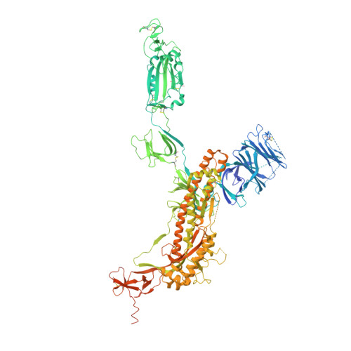
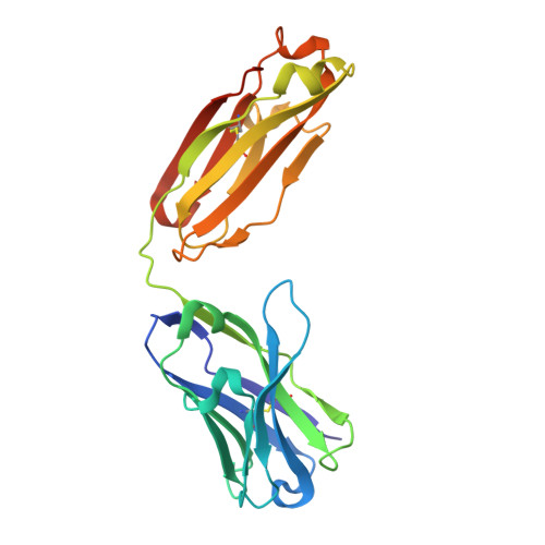
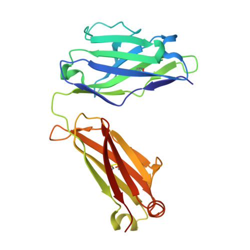
Mapping Neutralizing and Immunodominant Sites on the SARS-CoV-2 Spike Receptor-Binding Domain by Structure-Guided High-Resolution Serology.

Piccoli, L.Park, Y.J.Tortorici, M.A.Czudnochowski, N.Walls, A.C.Beltramello, M.Silacci-Fregni, C.Pinto, D.Rosen, L.E.Bowen, J.E.Acton, O.J.Jaconi, S.Guarino, B.Minola, A.Zatta, F.Sprugasci, N.Bassi, J.Peter, A.De Marco, A.Nix, J.C.Mele, F.Jovic, S.Rodriguez, B.F.Gupta, S.V.Jin, F.Piumatti, G.Lo Presti, G.Pellanda, A.F.Biggiogero, M.Tarkowski, M.Pizzuto, M.S.Cameroni, E.Havenar-Daughton, C.Smithey, M.Hong, D.Lepori, V.Albanese, E.Ceschi, A.Bernasconi, E.Elzi, L.Ferrari, P.Garzoni, C.Riva, A.Snell, G.Sallusto, F.Fink, K.Virgin, H.W.Lanzavecchia, A.Corti, D.Veesler, D.

(2020) Cell 183: 1024-1042.e21

  • DOI: https://doi.org/10.1016/j.cell.2020.09.037
  • Primary Citation Related Structures: 
    7JV2, 7JV4, 7JV6, 7JVA, 7JVC, 7JW0, 7JX3, 7JXC, 7JXD, 7JXE

  • PubMed Abstract: 

    Analysis of the specificity and kinetics of neutralizing antibodies (nAbs) elicited by SARS-CoV-2 infection is crucial for understanding immune protection and identifying targets for vaccine design. In a cohort of 647 SARS-CoV-2-infected subjects, we found that both the magnitude of Ab responses to SARS-CoV-2 spike (S) and nucleoprotein and nAb titers correlate with clinical scores. The receptor-binding domain (RBD) is immunodominant and the target of 90% of the neutralizing activity present in SARS-CoV-2 immune sera. Whereas overall RBD-specific serum IgG titers waned with a half-life of 49 days, nAb titers and avidity increased over time for some individuals, consistent with affinity maturation. We structurally defined an RBD antigenic map and serologically quantified serum Abs specific for distinct RBD epitopes leading to the identification of two major receptor-binding motif antigenic sites. Our results explain the immunodominance of the receptor-binding motif and will guide the design of COVID-19 vaccines and therapeutics.


  • Organizational Affiliation
    • Humabs BioMed SA, Vir Biotechnology, 6500 Bellinzona, Switzerland.

Macromolecule Content 

  • Total Structure Weight: 581.82 kDa 
  • Atom Count: 29,562 
  • Modeled Residue Count: 4,272 
  • Deposited Residue Count: 5,178 
  • Unique protein chains: 3

Macromolecules

Find similar proteins by:|  3D Structure
Entity ID: 1
MoleculeChains  Sequence LengthOrganismDetailsImage
Spike glycoproteinA,
D [auth B],
G [auth E]
1,281Severe acute respiratory syndrome coronavirus 2Mutation(s): 5 
Gene Names: S2
UniProt
Find proteins for P0DTC2 (Severe acute respiratory syndrome coronavirus 2)
Explore P0DTC2 
Go to UniProtKB:  P0DTC2
Entity Groups
Sequence Clusters30% Identity50% Identity70% Identity90% Identity95% Identity100% Identity
UniProt GroupP0DTC2
Glycosylation
Glycosylation Sites: 16Go to GlyGen: P0DTC2-1
Sequence Annotations
Expand
Reference Sequence
Find similar proteins by:|  3D Structure
Entity ID: 2
MoleculeChains  Sequence LengthOrganismDetailsImage
S2A4 Fab heavy chainB [auth H],
E [auth C],
H [auth F]
228Homo sapiensMutation(s): 0 
Find similar proteins by:|  3D Structure
Entity ID: 3
MoleculeChains  Sequence LengthOrganismDetailsImage
S2A4 Fab light chainC [auth L],
F [auth D],
I [auth G]
217Homo sapiensMutation(s): 0 

Oligosaccharides

Help  
Entity ID: 4
MoleculeChains Length2D Diagram GlycosylationD Interactions
2-acetamido-2-deoxy-beta-D-glucopyranose-(1-4)-2-acetamido-2-deoxy-beta-D-glucopyranose
J [auth I],
K [auth J],
M,
N,
O,
J [auth I],
K [auth J],
M,
N,
O,
P,
R,
S,
T,
U,
W,
X
2N-Glycosylation
Glycosylation Resources
GlyTouCan: G42666HT
GlyCosmos: G42666HT
GlyGen: G42666HT
Entity ID: 5
MoleculeChains Length2D Diagram GlycosylationD Interactions
beta-D-mannopyranose-(1-4)-2-acetamido-2-deoxy-beta-D-glucopyranose-(1-4)-2-acetamido-2-deoxy-beta-D-glucopyranoseL [auth K],
Q,
V
3N-Glycosylation
Glycosylation Resources
GlyTouCan: G15407YE
GlyCosmos: G15407YE
GlyGen: G15407YE

Small Molecules

Ligands 1 Unique
IDChains Name / Formula / InChI Key2D Diagram3D Interactions
NAG
(Subject of Investigation/LOI)

Query on NAG


Download:Ideal Coordinates CCD File
AA [auth A]
AB [auth E]
BA [auth A]
BB [auth E]
CA [auth A]
AA [auth A],
AB [auth E],
BA [auth A],
BB [auth E],
CA [auth A],
CB [auth E],
DA [auth A],
DB [auth E],
EA [auth A],
EB [auth E],
FA [auth A],
GA [auth A],
HA [auth A],
IA [auth A],
JA [auth B],
KA [auth B],
LA [auth B],
MA [auth B],
NA [auth B],
OA [auth B],
PA [auth B],
QA [auth B],
RA [auth B],
SA [auth B],
TA [auth B],
UA [auth E],
VA [auth E],
WA [auth E],
XA [auth E],
Y [auth A],
YA [auth E],
Z [auth A],
ZA [auth E]
2-acetamido-2-deoxy-beta-D-glucopyranose
C8 H15 N O6
OVRNDRQMDRJTHS-FMDGEEDCSA-N

Experimental Data & Validation

Experimental Data

  • Method: ELECTRON MICROSCOPY
  • Resolution: 3.30 Å
  • Aggregation State: PARTICLE 
  • Reconstruction Method: SINGLE PARTICLE 
EM Software:
TaskSoftware PackageVersion
RECONSTRUCTIONcryoSPARC

Structure Validation

View Full Validation Report


Entry History 

& Funding Information

Deposition Data


Funding OrganizationLocationGrant Number
National Institutes of Health/National Institute of General Medical Sciences (NIH/NIGMS)United StatesR01GM120553

Revision History  (Full details and data files)

  • Version 1.0: 2020-10-14
    Type: Initial release
  • Version 1.1: 2020-11-25
    Changes: Database references
  • Version 1.2: 2021-01-27
    Changes: Structure summary
  • Version 1.3: 2021-06-23
    Changes: Other, Structure summary
  • Version 1.4: 2024-10-23
    Changes: Data collection, Database references, Structure summary