7JTV

Structure of IMPa from Pseudomonas aeruginosa in complex with an O-glycopeptide


Experimental Data Snapshot

  • Method: X-RAY DIFFRACTION
  • Resolution: 2.45 Å
  • R-Value Free: 0.234 
  • R-Value Work: 0.180 
  • R-Value Observed: 0.183 

wwPDB Validation   3D Report Full Report


This is version 1.1 of the entry. See complete history


Literature

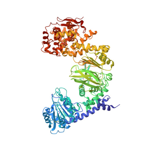
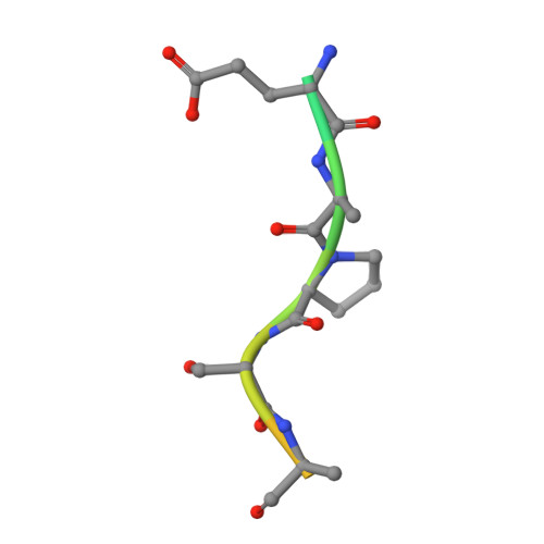
Structural evidence for a proline-specific glycopeptide recognition domain in an O-glycopeptidase.

Noach, I.Boraston, A.B.

(2021) Glycobiology 31: 385-390

  • DOI: https://doi.org/10.1093/glycob/cwaa095
  • Primary Citation of Related Structures:  
    7JTV

  • PubMed Abstract: 
  • The glycosylation of proteins is typically considered as a stabilizing modification, including resistance to proteolysis. A class of peptidases, referred to as glycopeptidases or O-glycopeptidases, circumvent the protective effect of glycans against proteolysis by accommodating the glycans in their active sites as specific features of substrate recognition ...

    The glycosylation of proteins is typically considered as a stabilizing modification, including resistance to proteolysis. A class of peptidases, referred to as glycopeptidases or O-glycopeptidases, circumvent the protective effect of glycans against proteolysis by accommodating the glycans in their active sites as specific features of substrate recognition. IMPa from Pseudomonas aeruginosa is such an O-glycopeptidase that cleaves the peptide bond immediately preceding a site of O-glycosylation, and through this glycoprotein-degrading function contributes to the host-pathogen interaction. IMPa, however, is a relatively large multidomain protein and how its additional domains may contribute to its function remains unknown. Here, through the determination of a crystal structure of IMPa in complex with an O-glycopeptide, we reveal that the N-terminal domain of IMPa, which is classified in Pfam as IMPa_N_2, is a proline recognition domain that also shows the properties of recognizing an O-linked glycan on the serine/threonine residue following the proline. The proline is bound in the center of a bowl formed by four functionally conserved aromatic amino acid side chains while the glycan wraps around one of the tyrosine residues in the bowl to make classic aromatic ring-carbohydrate CH-π interactions. This structural evidence provides unprecedented insight into how the ancillary domains in glycoprotein-specific peptidases can noncatalytically recognize specific glycosylated motifs that are common in mucin and mucin-like molecules.


    Organizational Affiliation

    Biochemistry & Microbiology, University of Victoria, PO Box 3055 STN CSC, Victoria, BC V8W 3P6, Canada.


Macromolecules
Find similar proteins by:  (by identity cutoff)  |  3D Structure
Entity ID: 1
MoleculeChainsSequence LengthOrganismDetailsImage
Immunomodulating metalloproteaseA [auth B],
B [auth A]
885Pseudomonas aeruginosa PAO1Mutation(s): 1 
Gene Names: impAPA0572
EC: 3.4.24
UniProt
Find proteins for Q9I5W4 (Pseudomonas aeruginosa (strain ATCC 15692 / DSM 22644 / CIP 104116 / JCM 14847 / LMG 12228 / 1C / PRS 101 / PAO1))
Explore Q9I5W4 
Go to UniProtKB:  Q9I5W4
Entity Groups  
Sequence Clusters30% Identity50% Identity70% Identity90% Identity95% Identity100% Identity
UniProt GroupQ9I5W4
Protein Feature View
Expand
  • Reference Sequence

Find similar proteins by:  Sequence   |   3D Structure  

Entity ID: 2
MoleculeChainsSequence LengthOrganismDetailsImage
GLU-ALA-PRO-SER-ALAC [auth E],
D [auth H]
15synthetic constructMutation(s): 0 
Entity Groups  
Sequence Clusters30% Identity50% Identity70% Identity90% Identity95% Identity100% Identity
Protein Feature View
Expand
  • Reference Sequence
Oligosaccharides

Help

Entity ID: 3
MoleculeChainsChain Length2D DiagramGlycosylation3D Interactions
beta-D-galactopyranose-(1-3)-2-acetamido-2-deoxy-alpha-D-galactopyranoseE [auth D],
F [auth G]
2O-Glycosylation Oligosaccharides Interaction
Glycosylation Resources
GlyTouCan:  G00031MO
GlyCosmos:  G00031MO
GlyGen:  G00031MO
Experimental Data & Validation

Experimental Data

  • Method: X-RAY DIFFRACTION
  • Resolution: 2.45 Å
  • R-Value Free: 0.234 
  • R-Value Work: 0.180 
  • R-Value Observed: 0.183 
  • Space Group: P 1 21 1
Unit Cell:
Length ( Å )Angle ( ˚ )
a = 90.34α = 90
b = 156.5β = 114.31
c = 95.68γ = 90
Software Package:
Software NamePurpose
PHENIXrefinement
XDSdata processing
Aimlessdata reduction
Aimlessdata scaling
PHASERphasing

Structure Validation

View Full Validation Report


Entry History & Funding Information

Deposition Data


Funding OrganizationLocationGrant Number
Canadian Institutes of Health Research (CIHR)Canada130305

Revision History  (Full details and data files)

  • Version 1.0: 2020-12-16
    Type: Initial release
  • Version 1.1: 2021-05-12
    Changes: Database references