7JO3

Carbonic Anhydrase IX Mimic Complexed with N-(5-sulfamoyl-1,3,4-thiadiazol-2-yl)pivalamide


Experimental Data Snapshot

  • Method: X-RAY DIFFRACTION
  • Resolution: 1.45 Å
  • R-Value Free: 0.173 
  • R-Value Work: 0.161 
  • R-Value Observed: 0.162 

wwPDB Validation   3D Report Full Report


Ligand Structure Quality Assessment 


This is version 1.2 of the entry. See complete history


Literature

Structural Basis of Nanomolar Inhibition of Tumor-Associated Carbonic Anhydrase IX: X-Ray Crystallographic and Inhibition Study of Lipophilic Inhibitors with Acetazolamide Backbone.

Andring, J.T.Fouch, M.Akocak, S.Angeli, A.Supuran, C.T.Ilies, M.A.McKenna, R.

(2020) J Med Chem 63: 13064-13075

  • DOI: https://doi.org/10.1021/acs.jmedchem.0c01390
  • Primary Citation of Related Structures:  
    7JNR, 7JNV, 7JNW, 7JNX, 7JNZ, 7JO0, 7JO1, 7JO2, 7JO3, 7JOB, 7JOC

  • PubMed Abstract: 

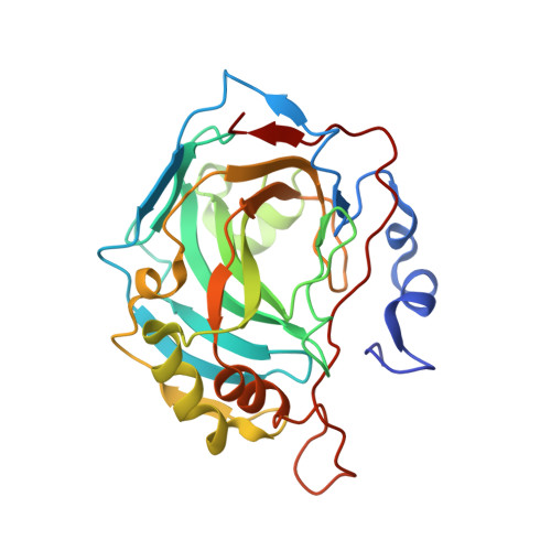
    This study provides a structure-activity relationship study of a series of lipophilic carbonic anhydrase (CA) inhibitors with an acetazolamide backbone. The inhibitors were tested against the tumor-expressed CA isozyme IX (CA IX), and the cytosolic CA I, CA II, and membrane-bound CA IV. The study identified several low nanomolar potent inhibitors against CA IX, with lipophilicities spanning two log units. Very potent pan-inhibitors with nanomolar potency against CA IX and sub-nanomolar potency against CA II and CA IV, and with potency against CA I one order of magnitude better than the parent acetazolamide 1 were also identified in this study, together with compounds that displayed selectivity against membrane-bound CA IV. A comprehensive X-ray crystallographic study (12 crystal structures), involving both CA II and a soluble CA IX mimetic (CA IX-mimic), revealed the structural basis of this particular inhibition profile and laid the foundation for further developments toward more potent and selective inhibitors for the tumor-expressed CA IX.


  • Organizational Affiliation

    Department of Biochemistry and Molecular Biology, College of Medicine, University of Florida, Gainesville, Florida 32610, United States.


Macromolecules
Find similar proteins by:  (by identity cutoff)  |  3D Structure
Entity ID: 1
MoleculeChains Sequence LengthOrganismDetailsImage
Carbonic anhydrase 2260Homo sapiensMutation(s): 7 
Gene Names: CA2
EC: 4.2.1.1
UniProt & NIH Common Fund Data Resources
Find proteins for P00918 (Homo sapiens)
Explore P00918 
Go to UniProtKB:  P00918
PHAROS:  P00918
GTEx:  ENSG00000104267 
Entity Groups  
Sequence Clusters30% Identity50% Identity70% Identity90% Identity95% Identity100% Identity
UniProt GroupP00918
Sequence Annotations
Expand
  • Reference Sequence
Small Molecules
Binding Affinity Annotations 
IDSourceBinding Affinity
VGS Binding MOAD:  7JO3 Ki: 8.2 (nM) from 1 assay(s)
Experimental Data & Validation

Experimental Data

  • Method: X-RAY DIFFRACTION
  • Resolution: 1.45 Å
  • R-Value Free: 0.173 
  • R-Value Work: 0.161 
  • R-Value Observed: 0.162 
  • Space Group: P 1 21 1
Unit Cell:
Length ( Å )Angle ( ˚ )
a = 41.652α = 90
b = 41.065β = 103.84
c = 71.431γ = 90
Software Package:
Software NamePurpose
PHENIXrefinement
Aimlessdata scaling
PDB_EXTRACTdata extraction
XDSdata reduction
PHENIXphasing

Structure Validation

View Full Validation Report


Ligand Structure Quality Assessment 


Entry History 

Deposition Data

Revision History  (Full details and data files)

  • Version 1.0: 2020-11-04
    Type: Initial release
  • Version 1.1: 2020-11-18
    Changes: Database references
  • Version 1.2: 2023-10-18
    Changes: Data collection, Database references, Refinement description