7JHP | pdb_00007jhp

Crystal structure of HRas in complex with the Ras-binding and cysteine-rich domains of CRaf-kinase


Experimental Data Snapshot

  • Method: X-RAY DIFFRACTION
  • Resolution: 2.77 Å
  • R-Value Free: 
    0.264 (Depositor), 0.262 (DCC) 
  • R-Value Work: 
    0.211 (Depositor), 0.212 (DCC) 
  • R-Value Observed: 
    0.216 (Depositor) 

Starting Models: experimental
View more details

wwPDB Validation 3D Report Full Report

Validation slider image for 7JHP

Ligand Structure Quality Assessment 


This is version 1.2 of the entry. See complete history

Literature

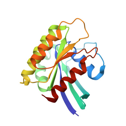
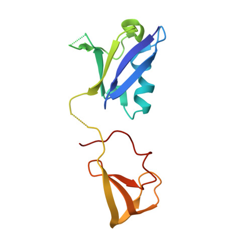
Crystal Structure Reveals the Full Ras-Raf Interface and Advances Mechanistic Understanding of Raf Activation.

Cookis, T.Mattos, C.

(2021) Biomolecules 11

  • DOI: https://doi.org/10.3390/biom11070996
  • Primary Citation Related Structures: 
    7JHP

  • PubMed Abstract: 

    Ras and Raf-kinase interact through the Ras-binding (RBD) and cysteine-rich domains (CRD) of Raf to signal through the mitogen-activated protein kinase pathway, yet the molecular mechanism leading to Raf activation has remained elusive. We present the 2.8 Å crystal structure of the HRas-CRaf-RBD_CRD complex showing the Ras-Raf interface as a continuous surface on Ras, as seen in the KRas-CRaf-RBD_CRD structure. In molecular dynamics simulations of a Ras dimer model formed through the α4-α5 interface, the CRD is dynamic and located between the two Ras protomers, poised for direct or allosteric modulation of functionally relevant regions of Ras and Raf. We propose a molecular model in which Ras binding is involved in the release of Raf autoinhibition while the Ras-Raf complex dimerizes to promote a platform for signal amplification, with Raf-CRD centrally located to impact regulation and function.


  • Organizational Affiliation
    • Department of Chemistry and Chemical Biology, Northeastern University, Boston, MA 02115, USA.

Macromolecule Content 

  • Total Structure Weight: 34.86 kDa 
  • Atom Count: 2,311 
  • Modeled Residue Count: 292 
  • Deposited Residue Count: 299 
  • Unique protein chains: 2

Macromolecules

Find similar proteins by:|  3D Structure
Entity ID: 1
MoleculeChains  Sequence LengthOrganismDetailsImage
GTPase HRas166Homo sapiensMutation(s): 1 
Gene Names: HRASHRAS1
EC: 3.6.5.2
UniProt & NIH Common Fund Data Resources
Find proteins for P01112 (Homo sapiens)
Explore P01112 
Go to UniProtKB:  P01112
PHAROS:  P01112
GTEx:  ENSG00000174775 
Entity Groups
Sequence Clusters30% Identity50% Identity70% Identity90% Identity95% Identity100% Identity
UniProt GroupP01112
Sequence Annotations
Expand
Reference Sequence
Find similar proteins by:|  3D Structure
Entity ID: 2
MoleculeChains  Sequence LengthOrganismDetailsImage
RAF proto-oncogene serine/threonine-protein kinaseB [auth C]133Homo sapiensMutation(s): 0 
Gene Names: RAF1RAF
EC: 2.7.11.1
UniProt & NIH Common Fund Data Resources
Find proteins for P04049 (Homo sapiens)
Explore P04049 
Go to UniProtKB:  P04049
PHAROS:  P04049
GTEx:  ENSG00000132155 
Entity Groups
Sequence Clusters30% Identity50% Identity70% Identity90% Identity95% Identity100% Identity
UniProt GroupP04049
Sequence Annotations
Expand
Reference Sequence

Experimental Data & Validation

Experimental Data

  • Method: X-RAY DIFFRACTION
  • Resolution: 2.77 Å
  • R-Value Free:  0.264 (Depositor), 0.262 (DCC) 
  • R-Value Work:  0.211 (Depositor), 0.212 (DCC) 
  • R-Value Observed: 0.216 (Depositor) 
Space Group: C 1 2 1
Unit Cell:
Length ( Å )Angle ( ˚ )
a = 94.487α = 90
b = 44.353β = 103.945
c = 74.395γ = 90
Software Package:
Software NamePurpose
PHENIXrefinement
PHENIXrefinement
HKL-3000data reduction
HKL-3000data scaling
PHENIXphasing

Structure Validation

View Full Validation Report


Ligand Structure Quality Assessment 


Entry History 

& Funding Information

Deposition Data


Funding OrganizationLocationGrant Number
National Science Foundation (NSF, United States)United StatesMCB-1517295

Revision History  (Full details and data files)

  • Version 1.0: 2020-08-05
    Type: Initial release
  • Version 1.1: 2021-08-25
    Changes: Database references
  • Version 1.2: 2023-10-18
    Changes: Data collection, Database references, Refinement description