7JHP

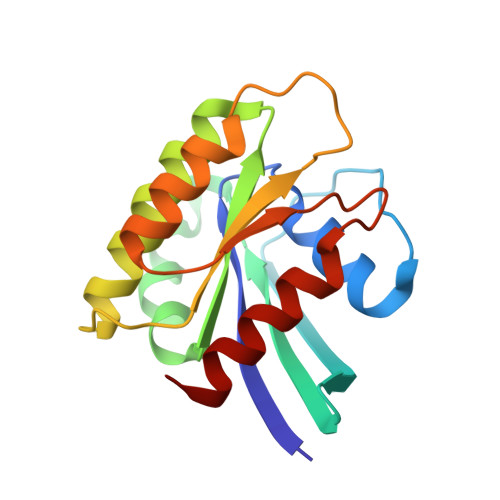
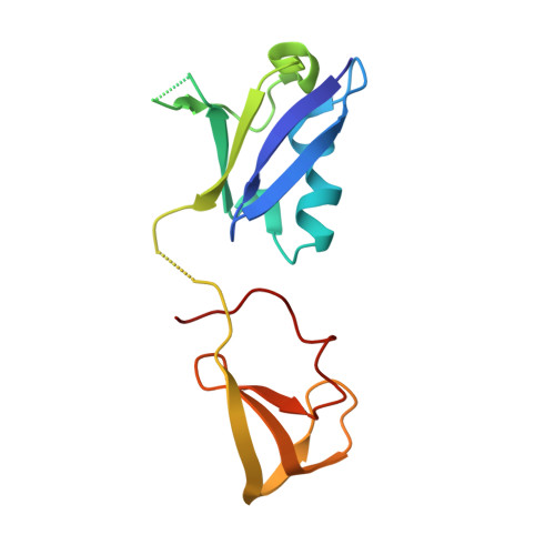
Crystal structure of HRas in complex with the Ras-binding and cysteine-rich domains of CRaf-kinase


Experimental Data Snapshot

  • Method: X-RAY DIFFRACTION
  • Resolution: 2.77 Å
  • R-Value Free: 0.264 
  • R-Value Work: 0.211 
  • R-Value Observed: 0.216 

wwPDB Validation   3D Report Full Report


Ligand Structure Quality Assessment 


This is version 1.2 of the entry. See complete history


Literature

Crystal Structure Reveals the Full Ras-Raf Interface and Advances Mechanistic Understanding of Raf Activation.

Cookis, T.Mattos, C.

(2021) Biomolecules 11

  • DOI: https://doi.org/10.3390/biom11070996
  • Primary Citation of Related Structures:  
    7JHP

  • PubMed Abstract: 

    Ras and Raf-kinase interact through the Ras-binding (RBD) and cysteine-rich domains (CRD) of Raf to signal through the mitogen-activated protein kinase pathway, yet the molecular mechanism leading to Raf activation has remained elusive. We present the 2.8 Å crystal structure of the HRas-CRaf-RBD_CRD complex showing the Ras-Raf interface as a continuous surface on Ras, as seen in the KRas-CRaf-RBD_CRD structure. In molecular dynamics simulations of a Ras dimer model formed through the α4-α5 interface, the CRD is dynamic and located between the two Ras protomers, poised for direct or allosteric modulation of functionally relevant regions of Ras and Raf. We propose a molecular model in which Ras binding is involved in the release of Raf autoinhibition while the Ras-Raf complex dimerizes to promote a platform for signal amplification, with Raf-CRD centrally located to impact regulation and function.


  • Organizational Affiliation

    Department of Chemistry and Chemical Biology, Northeastern University, Boston, MA 02115, USA.


Macromolecules
Find similar proteins by:  (by identity cutoff)  |  3D Structure
Entity ID: 1
MoleculeChains Sequence LengthOrganismDetailsImage
GTPase HRas166Homo sapiensMutation(s): 1 
Gene Names: HRASHRAS1
UniProt & NIH Common Fund Data Resources
Find proteins for P01112 (Homo sapiens)
Explore P01112 
Go to UniProtKB:  P01112
PHAROS:  P01112
GTEx:  ENSG00000174775 
Entity Groups  
Sequence Clusters30% Identity50% Identity70% Identity90% Identity95% Identity100% Identity
UniProt GroupP01112
Sequence Annotations
Expand
  • Reference Sequence
Find similar proteins by:  (by identity cutoff)  |  3D Structure
Entity ID: 2
MoleculeChains Sequence LengthOrganismDetailsImage
RAF proto-oncogene serine/threonine-protein kinaseB [auth C]133Homo sapiensMutation(s): 0 
Gene Names: RAF1RAF
EC: 2.7.11.1
UniProt & NIH Common Fund Data Resources
Find proteins for P04049 (Homo sapiens)
Explore P04049 
Go to UniProtKB:  P04049
PHAROS:  P04049
GTEx:  ENSG00000132155 
Entity Groups  
Sequence Clusters30% Identity50% Identity70% Identity90% Identity95% Identity100% Identity
UniProt GroupP04049
Sequence Annotations
Expand
  • Reference Sequence
Experimental Data & Validation

Experimental Data

  • Method: X-RAY DIFFRACTION
  • Resolution: 2.77 Å
  • R-Value Free: 0.264 
  • R-Value Work: 0.211 
  • R-Value Observed: 0.216 
  • Space Group: C 1 2 1
Unit Cell:
Length ( Å )Angle ( ˚ )
a = 94.487α = 90
b = 44.353β = 103.945
c = 74.395γ = 90
Software Package:
Software NamePurpose
PHENIXrefinement
PHENIXrefinement
HKL-3000data reduction
HKL-3000data scaling
PHENIXphasing

Structure Validation

View Full Validation Report


Ligand Structure Quality Assessment 


Entry History & Funding Information

Deposition Data


Funding OrganizationLocationGrant Number
National Science Foundation (NSF, United States)United StatesMCB-1517295

Revision History  (Full details and data files)

  • Version 1.0: 2020-08-05
    Type: Initial release
  • Version 1.1: 2021-08-25
    Changes: Database references
  • Version 1.2: 2023-10-18
    Changes: Data collection, Database references, Refinement description