Protonation states of hen egg-white lysozyme observed using D/H contrast neutron crystallography.
Chatake, T., Tanaka, I., Kusaka, K., Fujiwara, S.(2022) Acta Crystallogr D Struct Biol 78: 770-778
- PubMed: 35647923 Search on PubMed
- DOI: https://doi.org/10.1107/S2059798322004521
- Primary Citation Related Structures:
7FCU, 7FCW, 7VEI - PubMed Abstract:
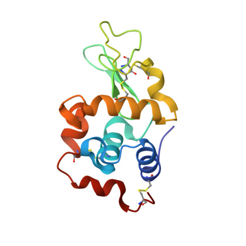
Hen egg-white lysozyme (HEWL) is an enzymatic protein with two acidic amino acids, Glu35 and Asp52, in its active site. Glu35 acts as a proton donor to the substrate and Asp52 interacts with the positively charged substrate, suggesting different protonation states of these residues. However, neutron crystallographic studies thus far have not provided a consistent picture of the protonation states of these residues. Only one study succeeded in observing the active protonation states of Glu35 and Asp52 in the triclinic crystal system. However, their active states in the most widely studied tetragonal crystal system are still unknown. The application of the D/H contrast technique in neutron crystallography improves the ability to locate exchangeable D/H atoms in proteins. In the present study, D 2 O and H 2 O solvent crystals were prepared. Each neutron data set was collected for only five days by combining a time-of-flight diffractometer (iBIX) and the spallation neutron source at the Japan Proton Accelerator Research Complex. The D/H contrast map provided better visualization of the D/H atoms in HEWL than the conventional neutron scattering length density map. The neutron D/H contrast map demonstrated the alternative protonation of the OE1 and OE2 atoms in the carboxyl group of Glu35. This alternative protonation occurs in the absence of a substrate, where high selectivity of the protonation site does not occur. In this case, only the OE1-HE1 bond attacks the substrate in an equilibrium between OE1-HE1 and OE2-HE2, or the H + ion of the OE2-HE2 bond moves to the OE1 atom just before or after substrate binding to initiate the catalytic reaction. In contrast, the carboxyl group of Asp52 is not protonated. Protonation of the carboxyl group was not observed for other Asp and Glu residues. These results are consistent with results from NMR spectroscopy and explain the protonation states at the active site in the apo form of HEWL.
- Institute for Integrated Radiation and Nuclear Science, Kyoto University, Asashironishi 2, Kumatori, Osaka 590-0494, Japan.
Organizational Affiliation: