7F6Z | pdb_00007f6z

Complex Structure of antibody BD-503 and RBD-501Y.V2 of COVID-19


Experimental Data Snapshot

  • Method: X-RAY DIFFRACTION
  • Resolution: 3.00 Å
  • R-Value Free: 
    0.254 (Depositor), 0.254 (DCC) 
  • R-Value Work: 
    0.207 (Depositor), 0.209 (DCC) 
  • R-Value Observed: 
    0.211 (Depositor) 

Starting Model: experimental
View more details

wwPDB Validation 3D Report Full Report

Validation slider image for 7F6Z

This is version 1.3 of the entry. See complete history

Literature

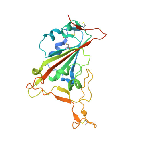
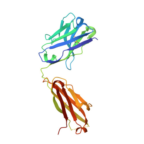
Structure-based analyses of neutralization antibodies interacting with naturally occurring SARS-CoV-2 RBD variants.

Xu, H.Wang, B.Zhao, T.N.Liang, Z.T.Peng, T.B.Song, X.H.Wu, J.J.Wang, Y.C.Su, X.D.

(2021) Cell Res 31: 1126-1129

  • DOI: https://doi.org/10.1038/s41422-021-00554-1
  • Primary Citation Related Structures: 
    7EJY, 7EJZ, 7EK0, 7F6Y, 7F6Z

  • Organizational Affiliation
    • State Key Laboratory of Protein and Plant Gene Research, School of Life Sciences, and Biomedical Pioneering Innovation Center (BIOPIC), Peking University, Beijing, China.

Macromolecule Content 

  • Total Structure Weight: 72.21 kDa 
  • Atom Count: 4,780 
  • Modeled Residue Count: 622 
  • Deposited Residue Count: 660 
  • Unique protein chains: 3

Macromolecules

Find similar proteins by:|  3D Structure
Entity ID: 1
MoleculeChains  Sequence LengthOrganismDetailsImage
Spike protein S1A [auth R]223Severe acute respiratory syndrome coronavirus 2Mutation(s): 3 
Gene Names: S2
UniProt
Find proteins for P0DTC2 (Severe acute respiratory syndrome coronavirus 2)
Explore P0DTC2 
Go to UniProtKB:  P0DTC2
Entity Groups
Sequence Clusters30% Identity50% Identity70% Identity90% Identity95% Identity100% Identity
UniProt GroupP0DTC2
Glycosylation
Glycosylation Sites: 1Go to GlyGen: P0DTC2-1
Sequence Annotations
Expand
Reference Sequence
Find similar proteins by:|  3D Structure
Entity ID: 2
MoleculeChains  Sequence LengthOrganismDetailsImage
Heavy Chain of BD-503B [auth H]222Homo sapiensMutation(s): 0 
Find similar proteins by:|  3D Structure
Entity ID: 3
MoleculeChains  Sequence LengthOrganismDetailsImage
Light Chain of BD-503C [auth L]215Homo sapiensMutation(s): 0 

Oligosaccharides

Help  
Entity ID: 4
MoleculeChains Length2D Diagram GlycosylationD Interactions
2-acetamido-2-deoxy-beta-D-glucopyranose-(1-4)-2-acetamido-2-deoxy-beta-D-glucopyranoseD [auth A]2N-Glycosylation
Glycosylation Resources
GlyTouCan: G42666HT
GlyCosmos: G42666HT
GlyGen: G42666HT

Experimental Data & Validation

Experimental Data

  • Method: X-RAY DIFFRACTION
  • Resolution: 3.00 Å
  • R-Value Free:  0.254 (Depositor), 0.254 (DCC) 
  • R-Value Work:  0.207 (Depositor), 0.209 (DCC) 
  • R-Value Observed: 0.211 (Depositor) 
Space Group: C 2 2 21
Unit Cell:
Length ( Å )Angle ( ˚ )
a = 82.92α = 90
b = 146.837β = 90
c = 145.651γ = 90
Software Package:
Software NamePurpose
PHENIXrefinement
XDSdata reduction
XSCALEdata scaling
PHASERphasing

Structure Validation

View Full Validation Report


Entry History 

Deposition Data

Revision History  (Full details and data files)

  • Version 1.0: 2022-06-29
    Type: Initial release
  • Version 1.1: 2022-11-02
    Changes: Database references
  • Version 1.2: 2023-11-29
    Changes: Data collection, Refinement description
  • Version 1.3: 2024-11-13
    Changes: Structure summary