7F5H

The crystal structure of RBD-Nanobody complex, DL28 (SC4)


Experimental Data Snapshot

  • Method: X-RAY DIFFRACTION
  • Resolution: 3.00 Å
  • R-Value Free: 0.248 
  • R-Value Work: 0.226 
  • R-Value Observed: 0.228 

wwPDB Validation   3D Report Full Report


This is version 1.2 of the entry. See complete history


Literature

Structural Characterization of a Neutralizing Nanobody With Broad Activity Against SARS-CoV-2 Variants.

Li, T.Zhou, B.Luo, Z.Lai, Y.Huang, S.Zhou, Y.Li, Y.Gautam, A.Bourgeau, S.Wang, S.Bao, J.Tan, J.Lavillette, D.Li, D.

(2022) Front Microbiol 13: 875840-875840

  • DOI: https://doi.org/10.3389/fmicb.2022.875840
  • Primary Citation of Related Structures:  
    7F5H

  • PubMed Abstract: 

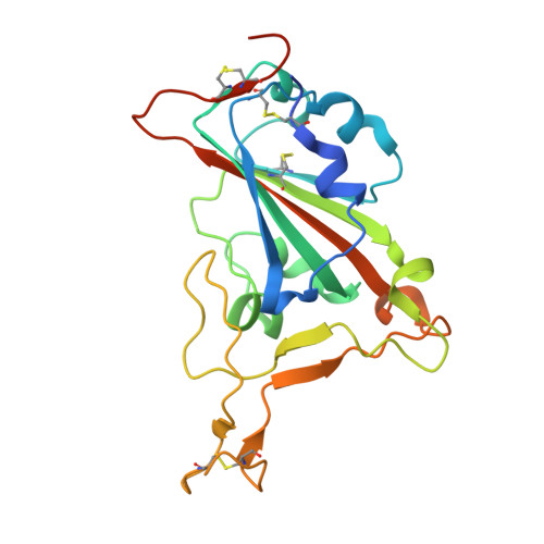
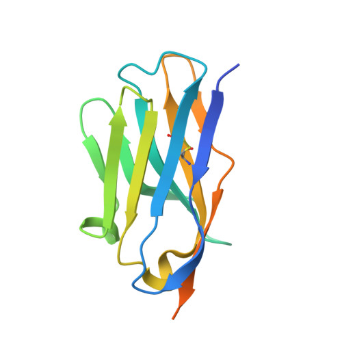
    SARS-CoV-2 and its variants, such as the Omicron continue to threaten public health. The virus recognizes the host cell by attaching its Spike (S) receptor-binding domain (RBD) to the host receptor, ACE2. Therefore, RBD is a primary target for neutralizing antibodies and vaccines. Here, we report the isolation and biological and structural characterization of a single-chain antibody (nanobody) from RBD-immunized alpaca. The nanobody, named DL28, binds to RBD tightly with a K D of 1.56 nM and neutralizes the original SARS-CoV-2 strain with an IC 50 of 0.41 μg mL -1 . Neutralization assays with a panel of variants of concern (VOCs) reveal its wide-spectrum activity with IC 50 values ranging from 0.35 to 1.66 μg mL -1 for the Alpha/Beta/Gamma/Delta and an IC 50 of 0.66 μg mL -1 for the currently prevalent Omicron. Competition binding assays show that DL28 blocks ACE2-binding. However, structural characterizations and mutagenesis suggest that unlike most antibodies, the blockage by DL28 does not involve direct competition or steric hindrance. Rather, DL28 may use a "conformation competition" mechanism where it excludes ACE2 by keeping an RBD loop in a conformation incompatible with ACE2-binding.


  • Organizational Affiliation

    State Key Laboratory of Molecular Biology, Chinese Academy of Sciences Center for Excellence in Molecular Cell Science, Shanghai Institute of Biochemistry and Cell Biology, Chinese Academy of Sciences, Shanghai, China.


Macromolecules
Find similar proteins by:  (by identity cutoff)  |  3D Structure
Entity ID: 1
MoleculeChains Sequence LengthOrganismDetailsImage
SARS-CoV-2 Spike Receptor-Binding Domain (RBD)
A, B
213Severe acute respiratory syndrome coronavirus 2Mutation(s): 0 
Gene Names: S2
UniProt
Find proteins for P0DTC2 (Severe acute respiratory syndrome coronavirus 2)
Explore P0DTC2 
Go to UniProtKB:  P0DTC2
Entity Groups  
Sequence Clusters30% Identity50% Identity70% Identity90% Identity95% Identity100% Identity
UniProt GroupP0DTC2
Sequence Annotations
Expand
  • Reference Sequence
Find similar proteins by:  (by identity cutoff)  |  3D Structure
Entity ID: 2
MoleculeChains Sequence LengthOrganismDetailsImage
Nanobody DL28
C, D
143Vicugna pacosMutation(s): 0 
Entity Groups  
Sequence Clusters30% Identity50% Identity70% Identity90% Identity95% Identity100% Identity
Sequence Annotations
Expand
  • Reference Sequence
Oligosaccharides

Help

Entity ID: 3
MoleculeChains Length2D Diagram Glycosylation3D Interactions
beta-D-mannopyranose-(1-4)-2-acetamido-2-deoxy-beta-D-glucopyranose-(1-4)-[alpha-L-fucopyranose-(1-6)]2-acetamido-2-deoxy-beta-D-glucopyranose
E, F
4N-Glycosylation
Glycosylation Resources
GlyTouCan:  G32152BH
GlyCosmos:  G32152BH
GlyGen:  G32152BH
Experimental Data & Validation

Experimental Data

  • Method: X-RAY DIFFRACTION
  • Resolution: 3.00 Å
  • R-Value Free: 0.248 
  • R-Value Work: 0.226 
  • R-Value Observed: 0.228 
  • Space Group: P 65 2 2
Unit Cell:
Length ( Å )Angle ( ˚ )
a = 177.463α = 90
b = 177.463β = 90
c = 133.13γ = 120
Software Package:
Software NamePurpose
REFMACrefinement
HKL-2000data reduction
HKL-2000data scaling
PHASERphasing

Structure Validation

View Full Validation Report


Entry History & Funding Information

Deposition Data


Funding OrganizationLocationGrant Number
National Natural Science Foundation of China (NSFC)China31870726

Revision History  (Full details and data files)

  • Version 1.0: 2022-06-29
    Type: Initial release
  • Version 1.1: 2023-01-11
    Changes: Database references
  • Version 1.2: 2023-11-29
    Changes: Data collection, Refinement description