7F2X

Crystal structure of MEK1 C121S mutant


Experimental Data Snapshot

  • Method: X-RAY DIFFRACTION
  • Resolution: 2.01 Å
  • R-Value Free: 0.232 
  • R-Value Work: 0.215 
  • R-Value Observed: 0.216 

wwPDB Validation   3D Report Full Report


Ligand Structure Quality Assessment 


This is version 1.2 of the entry. See complete history


Literature

Qualitative differences in disease-associated MEK mutants reveal molecular signatures and aberrant signaling-crosstalk in cancer.

Kubota, Y.Fujioka, Y.Patil, A.Takagi, Y.Matsubara, D.Iijima, M.Momose, I.Naka, R.Nakai, K.Noda, N.N.Takekawa, M.

(2022) Nat Commun 13: 4063-4063

  • DOI: https://doi.org/10.1038/s41467-022-31690-w
  • Primary Citation of Related Structures:  
    7F2X

  • PubMed Abstract: 

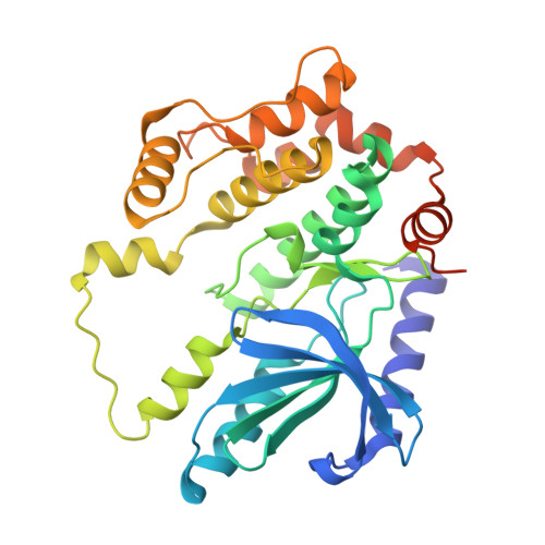
    Point-mutations of MEK1, a central component of ERK signaling, are present in cancer and RASopathies, but their precise biological effects remain obscure. Here, we report a mutant MEK1 structure that uncovers the mechanisms underlying abnormal activities of cancer- and RASopathy-associated MEK1 mutants. These two classes of MEK1 mutations differentially impact on spatiotemporal dynamics of ERK signaling, cellular transcriptional programs, gene expression profiles, and consequent biological outcomes. By making use of such distinct characteristics of the MEK1 mutants, we identified cancer- and RASopathy-signature genes that may serve as diagnostic markers or therapeutic targets for these diseases. In particular, two AKT-inhibitor molecules, PHLDA1 and 2, are simultaneously upregulated by oncogenic ERK signaling, and mediate cancer-specific ERK-AKT crosstalk. The combined expression of PHLDA1/2 is critical to confer resistance to ERK pathway-targeted therapeutics on cancer cells. Finally, we propose a therapeutic strategy to overcome this drug resistance. Our data provide vital insights into the etiology, diagnosis, and therapeutic strategy of cancers and RASopathies.


  • Organizational Affiliation

    Division of Cell Signaling and Molecular Medicine, Institute of Medical Science, The University of Tokyo, Minato-ku, Tokyo, 108-8639, Japan.


Macromolecules
Find similar proteins by:  (by identity cutoff)  |  3D Structure
Entity ID: 1
MoleculeChains Sequence LengthOrganismDetailsImage
MEK1 F11321Homo sapiensMutation(s): 0 
UniProt & NIH Common Fund Data Resources
Find proteins for Q02750 (Homo sapiens)
Explore Q02750 
Go to UniProtKB:  Q02750
PHAROS:  Q02750
GTEx:  ENSG00000169032 
Entity Groups  
Sequence Clusters30% Identity50% Identity70% Identity90% Identity95% Identity100% Identity
UniProt GroupQ02750
Sequence Annotations
Expand
  • Reference Sequence
Small Molecules
Ligands 1 Unique
IDChains Name / Formula / InChI Key2D Diagram3D Interactions
ANP (Subject of Investigation/LOI)
Query on ANP

Download Ideal Coordinates CCD File 
B [auth A]PHOSPHOAMINOPHOSPHONIC ACID-ADENYLATE ESTER
C10 H17 N6 O12 P3
PVKSNHVPLWYQGJ-KQYNXXCUSA-N
Experimental Data & Validation

Experimental Data

  • Method: X-RAY DIFFRACTION
  • Resolution: 2.01 Å
  • R-Value Free: 0.232 
  • R-Value Work: 0.215 
  • R-Value Observed: 0.216 
  • Space Group: F 2 2 2
Unit Cell:
Length ( Å )Angle ( ˚ )
a = 49.498α = 90
b = 156.504β = 90
c = 179.156γ = 90
Software Package:
Software NamePurpose
PHENIXrefinement
XDSdata reduction
XDSdata scaling
BALBESphasing

Structure Validation

View Full Validation Report


Ligand Structure Quality Assessment 


Entry History 

Deposition Data

Revision History  (Full details and data files)

  • Version 1.0: 2022-06-22
    Type: Initial release
  • Version 1.1: 2022-08-10
    Changes: Database references
  • Version 1.2: 2023-11-29
    Changes: Data collection, Refinement description