7EG2 | pdb_00007eg2

Crystal structure of the apoAequorin complex with (S)-daCTZ


Experimental Data Snapshot

  • Method: X-RAY DIFFRACTION
  • Resolution: 2.22 Å
  • R-Value Free: 
    0.245 (Depositor), 0.245 (DCC) 
  • R-Value Work: 
    0.205 (Depositor), 0.206 (DCC) 
  • R-Value Observed: 
    0.205 (Depositor) 

Starting Model: experimental
View more details

wwPDB Validation 3D Report Full Report

Validation slider image for 7EG2

Ligand Structure Quality Assessment 


This is version 1.1 of the entry. See complete history

Literature

Chiral deaza-coelenterazine analogs for probing a substrate-binding site in the Ca2+-binding photoprotein aequorin.

Inouye, S.Sumida, Y.Tomabechi, Y.Taguchi, J.Shirouzu, M.Hosoya, T.

(2021) PLoS One 16: e0251743-e0251743

  • DOI: https://doi.org/10.1371/journal.pone.0251743
  • Primary Citation Related Structures: 
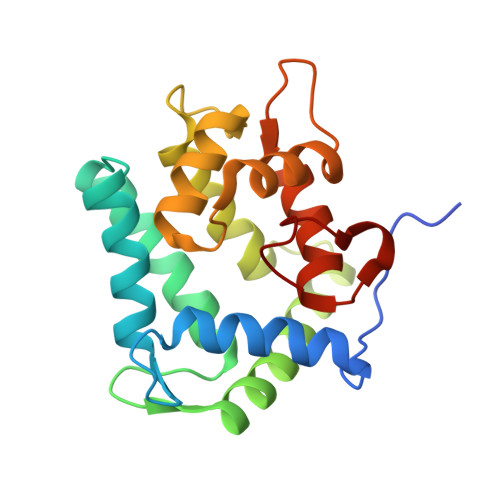
    7EG2, 7EG3

  • PubMed Abstract: 

    The Ca2+-binding photoprotein aequorin is a complex of apoAequorin (apoprotein) and (S)-2-peroxycoelenterazine. Aequorin can be regenerated by the incubation of apoAequorin with coelenterazine and molecular oxygen (O2). In this study, to investigate the molecular recognition of apoAequorin for coelenterazine using chemical probes, the chiral deaza-analogs of (S)- and (R)-deaza-CTZ (daCTZ) for coelenterazine and of (S)-2- and (R)-2-hydroxymethyl-deaza-CTZ (HM-daCTZ) for 2-peroxycoelenterazine were efficiently prepared by the improvement method. The chiral deaza-analogs of (S)-daCTZ and (S)-HM-daCTZ selectively inhibited the regeneration step to aequorin by binding the catalytic site of coelenterazine in the apoAequorin molecule. The crystal structures of the apoAequorin complexes with (S)-daCTZ and (S)-HM-daCTZ were determined, suggesting that the hydroxy moiety at the C6-hydroxyphenyl group and the carbonyl moiety of the imidazopyrazinone ring in coelenterazine are essential to bind the apoAequorin molecule through hydrogen bonding. Therefore, the chiral deaza-analogs of coelenterazine can be used as a probe to study the interaction between coelenterazine and the related proteins including photoprotein, luciferase, and coelenterazine-binding protein.


  • Organizational Affiliation
    • Yokohama Research Center, JNC Co., Yokohama, Japan.

Macromolecule Content 

  • Total Structure Weight: 367.91 kDa 
  • Atom Count: 26,432 
  • Modeled Residue Count: 3,051 
  • Deposited Residue Count: 3,168 
  • Unique protein chains: 1

Macromolecules

Find similar proteins by:|  3D Structure
Entity ID: 1
MoleculeChains  Sequence LengthOrganismDetailsImage
Aequorin-2
A, B, C, D, E
A, B, C, D, E, F, G, H, I, J, K, L, M, N, O, P
198Aequorea victoriaMutation(s): 0 
UniProt
Find proteins for P02592 (Aequorea victoria)
Explore P02592 
Go to UniProtKB:  P02592
Entity Groups
Sequence Clusters30% Identity50% Identity70% Identity90% Identity95% Identity100% Identity
UniProt GroupP02592
Sequence Annotations
Expand
Reference Sequence

Small Molecules

Ligands 1 Unique
IDChains Name / Formula / InChI Key2D Diagram3D Interactions
J2X
(Subject of Investigation/LOI)

Query on J2X


Download:Ideal Coordinates CCD File
AA [auth K]
BA [auth L]
CA [auth M]
DA [auth N]
EA [auth O]
AA [auth K],
BA [auth L],
CA [auth M],
DA [auth N],
EA [auth O],
FA [auth P],
Q [auth A],
R [auth B],
S [auth C],
T [auth D],
U [auth E],
V [auth F],
W [auth G],
X [auth H],
Y [auth I],
Z [auth J]
(2~{S})-2-(hydroxymethyl)-6-(4-hydroxyphenyl)-2-[(4-hydroxyphenyl)methyl]-4-(phenylmethyl)-3~{H}-inden-1-one
C30 H26 O4
OCWYWPXFECTDDD-PMERELPUSA-N

Experimental Data & Validation

Experimental Data

  • Method: X-RAY DIFFRACTION
  • Resolution: 2.22 Å
  • R-Value Free:  0.245 (Depositor), 0.245 (DCC) 
  • R-Value Work:  0.205 (Depositor), 0.206 (DCC) 
  • R-Value Observed: 0.205 (Depositor) 
Space Group: P 1
Unit Cell:
Length ( Å )Angle ( ˚ )
a = 91.31α = 77.47
b = 98.14β = 73.47
c = 121.48γ = 75.16
Software Package:
Software NamePurpose
PHASERphasing
PHENIXrefinement
PDB_EXTRACTdata extraction
XDSdata reduction
XDSdata scaling

Structure Validation

View Full Validation Report


Ligand Structure Quality Assessment 


Entry History 

Deposition Data

Revision History  (Full details and data files)

  • Version 1.0: 2021-06-23
    Type: Initial release
  • Version 1.1: 2023-11-29
    Changes: Data collection, Database references, Refinement description