7E40

Mechanism of Phosphate Sensing and Signaling Revealed by Rice SPX1-PHR2 Complex Structure


Experimental Data Snapshot

  • Method: X-RAY DIFFRACTION
  • Resolution: 2.60 Å
  • R-Value Free: 0.274 
  • R-Value Work: 0.226 
  • R-Value Observed: 0.228 

wwPDB Validation   3D Report Full Report


Ligand Structure Quality Assessment 


This is version 1.1 of the entry. See complete history


Literature

Mechanism of phosphate sensing and signaling revealed by rice SPX1-PHR2 complex structure.

Zhou, J.Hu, Q.Xiao, X.Yao, D.Ge, S.Ye, J.Li, H.Cai, R.Liu, R.Meng, F.Wang, C.Zhu, J.K.Lei, M.Xing, W.

(2021) Nat Commun 12: 7040-7040

  • DOI: https://doi.org/10.1038/s41467-021-27391-5
  • Primary Citation of Related Structures:  
    7E40

  • PubMed Abstract: 

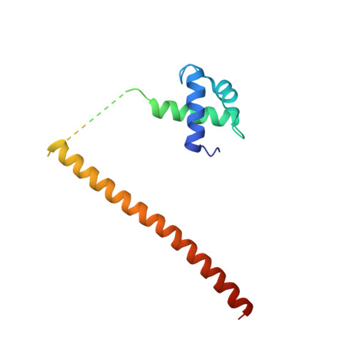
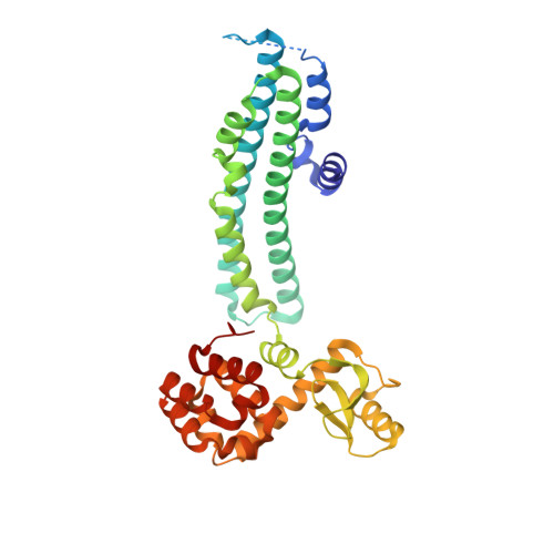
    Phosphate, a key plant nutrient, is perceived through inositol polyphosphates (InsPs) by SPX domain-containing proteins. SPX1 an inhibit the PHR2 transcription factor to maintain Pi homeostasis. How SPX1 recognizes an InsP molecule and represses transcription activation by PHR2 remains unclear. Here we show that, upon binding InsP 6 , SPX1 can disrupt PHR2 dimers and form a 1:1 SPX1-PHR2 complex. The complex structure reveals that SPX1 helix α1 can impose a steric hindrance when interacting with the PHR2 dimer. By stabilizing helix α1, InsP 6 allosterically decouples the PHR2 dimer and stabilizes the SPX1-PHR2 interaction. In doing so, InsP 6 further allows SPX1 to engage with the PHR2 MYB domain and sterically block its interaction with DNA. Taken together, our results suggest that, upon sensing the surrogate signals of phosphate, SPX1 inhibits PHR2 via a dual mechanism that attenuates dimerization and DNA binding activities of PHR2.


  • Organizational Affiliation

    Shanghai Center for Plant Stress Biology and Center of Excellence in Molecular Plant Sciences, Chinese Academy of Sciences, Shanghai, 200032, China.


Macromolecules
Find similar proteins by:  (by identity cutoff)  |  3D Structure
Entity ID: 1
MoleculeChains Sequence LengthOrganismDetailsImage
Protein PHOSPHATE STARVATION RESPONSE 2
A, C
134Oryza sativa Japonica GroupMutation(s): 4 
Gene Names: PHR2Os07g0438800LOC_Os07g25710OSJNBa0026I22.19P0443H10.4
UniProt
Find proteins for Q6Z156 (Oryza sativa subsp. japonica)
Explore Q6Z156 
Go to UniProtKB:  Q6Z156
Entity Groups  
Sequence Clusters30% Identity50% Identity70% Identity90% Identity95% Identity100% Identity
UniProt GroupQ6Z156
Sequence Annotations
Expand
  • Reference Sequence
Find similar proteins by:  (by identity cutoff)  |  3D Structure
Entity ID: 2
MoleculeChains Sequence LengthOrganismDetailsImage
SPX domain-containing protein 1,Endolysin
B, D
358Oryza sativa Japonica GroupTequatrovirus T4
This entity is chimeric
Mutation(s): 2 
Gene Names: SPX1Os06g0603600LOC_Os06g40120OsJ_21901P0486H12.37eT4Tp126
EC: 3.2.1.17
UniProt
Find proteins for Q69XJ0 (Oryza sativa subsp. japonica)
Explore Q69XJ0 
Go to UniProtKB:  Q69XJ0
Find proteins for D9IEF7 (Enterobacteria phage T4)
Explore D9IEF7 
Go to UniProtKB:  D9IEF7
Entity Groups  
Sequence Clusters30% Identity50% Identity70% Identity90% Identity95% Identity100% Identity
UniProt GroupsD9IEF7Q69XJ0
Sequence Annotations
Expand
  • Reference Sequence
Experimental Data & Validation

Experimental Data

  • Method: X-RAY DIFFRACTION
  • Resolution: 2.60 Å
  • R-Value Free: 0.274 
  • R-Value Work: 0.226 
  • R-Value Observed: 0.228 
  • Space Group: P 21 21 21
Unit Cell:
Length ( Å )Angle ( ˚ )
a = 65.244α = 90
b = 107.492β = 90
c = 174.521γ = 90
Software Package:
Software NamePurpose
PHENIXrefinement
XDSdata scaling
PDB_EXTRACTdata extraction
XDSdata reduction
PHENIXphasing

Structure Validation

View Full Validation Report


Ligand Structure Quality Assessment 


Entry History 

Deposition Data

Revision History  (Full details and data files)

  • Version 1.0: 2021-11-10
    Type: Initial release
  • Version 1.1: 2021-12-15
    Changes: Database references