7DZK

Fabp protein after hv


Experimental Data Snapshot

  • Method: X-RAY DIFFRACTION
  • Resolution: 1.54 Å
  • R-Value Free: 0.223 
  • R-Value Work: 0.191 
  • R-Value Observed: 0.192 

wwPDB Validation   3D Report Full Report


Ligand Structure Quality Assessment 


This is version 1.3 of the entry. See complete history


Literature

Excited-state intermediates in a designer protein encoding a phototrigger caught by an X-ray free-electron laser.

Liu, X.Liu, P.Li, H.Xu, Z.Jia, L.Xia, Y.Yu, M.Tang, W.Zhu, X.Chen, C.Zhang, Y.Nango, E.Tanaka, R.Luo, F.Kato, K.Nakajima, Y.Kishi, S.Yu, H.Matsubara, N.Owada, S.Tono, K.Iwata, S.Yu, L.J.Shen, J.R.Wang, J.

(2022) Nat Chem 14: 1054-1060

  • DOI: https://doi.org/10.1038/s41557-022-00992-3
  • Primary Citation of Related Structures:  
    7DZE, 7DZF, 7DZG, 7DZH, 7DZI, 7DZJ, 7DZK, 7DZL

  • PubMed Abstract: 

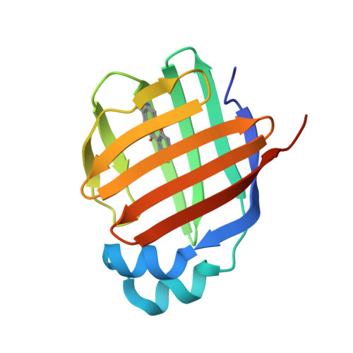
    One of the primary objectives in chemistry research is to observe atomic motions during reactions in real time. Although X-ray free-electron lasers (XFELs) have facilitated the capture of reaction intermediates using time-resolved serial femtosecond crystallography (TR-SFX), only a few natural photoactive proteins have been investigated using this method, mostly due to the lack of suitable phototriggers. Here we report the genetic encoding of a xanthone amino acid (FXO), as an efficient phototrigger, into a rationally designed human liver fatty-acid binding protein mutant (termed XOM), which undergoes photo-induced C-H bond transformation with high selectivity and quantum efficiency. We solved the structures of XOM before and 10-300 ns after flash illumination, at 1.55-1.70 Å resolutions, and captured the elusive excited-state intermediates responsible for precise C-H bond activation. We expect that most redox enzymes can now be investigated by TR-SFX, using our method, to reveal reaction intermediates key for their efficiency and selectivity.


  • Organizational Affiliation

    Laboratory of RNA Biology, Institute of Biophysics, Chinese Academy of Sciences, University of Chinese Academy of Sciences, Beijing, China.


Macromolecules
Find similar proteins by:  (by identity cutoff)  |  3D Structure
Entity ID: 1
MoleculeChains Sequence LengthOrganismDetailsImage
Fatty acid-binding protein, liver
A, B
137Homo sapiensMutation(s): 2 
Gene Names: FABP1FABPL
UniProt & NIH Common Fund Data Resources
Find proteins for P07148 (Homo sapiens)
Explore P07148 
Go to UniProtKB:  P07148
PHAROS:  P07148
GTEx:  ENSG00000163586 
Entity Groups  
Sequence Clusters30% Identity50% Identity70% Identity90% Identity95% Identity100% Identity
UniProt GroupP07148
Sequence Annotations
Expand
  • Reference Sequence
Small Molecules
Modified Residues  1 Unique
IDChains TypeFormula2D DiagramParent
HP9
Query on HP9
A, B
L-PEPTIDE LINKINGC16 H12 F N O4PHE
Experimental Data & Validation

Experimental Data

  • Method: X-RAY DIFFRACTION
  • Resolution: 1.54 Å
  • R-Value Free: 0.223 
  • R-Value Work: 0.191 
  • R-Value Observed: 0.192 
  • Space Group: P 21 21 21
Unit Cell:
Length ( Å )Angle ( ˚ )
a = 56.922α = 90
b = 66.157β = 90
c = 68.136γ = 90
Software Package:
Software NamePurpose
PHENIXrefinement
PDB_EXTRACTdata extraction
HKL-2000data reduction
HKL-2000data scaling
PHASERphasing

Structure Validation

View Full Validation Report


Ligand Structure Quality Assessment 


Entry History & Funding Information

Deposition Data


Funding OrganizationLocationGrant Number
National Natural Science Foundation of China (NSFC)China--

Revision History  (Full details and data files)

  • Version 1.0: 2022-07-27
    Type: Initial release
  • Version 1.1: 2022-08-17
    Changes: Database references
  • Version 1.2: 2022-09-07
    Changes: Database references
  • Version 1.3: 2023-11-29
    Changes: Data collection, Refinement description