7DY1

Crystal Structure of Cyanobacterial Circadian Clock Protein KaiC


Experimental Data Snapshot

  • Method: X-RAY DIFFRACTION
  • Resolution: 2.20 Å
  • R-Value Free: 0.284 
  • R-Value Work: 0.233 
  • R-Value Observed: 0.235 

wwPDB Validation   3D Report Full Report


Ligand Structure Quality Assessment 


This is version 1.1 of the entry. See complete history


Literature

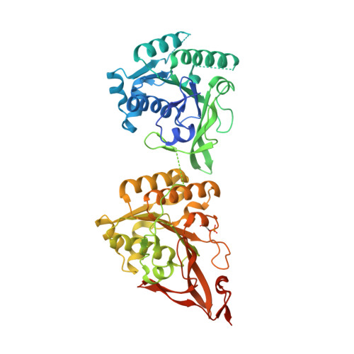
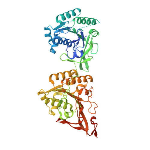
Regulation mechanisms of the dual ATPase in KaiC.

Furuike, Y.Mukaiyama, A.Koda, S.I.Simon, D.Ouyang, D.Ito-Miwa, K.Saito, S.Yamashita, E.Nishiwaki-Ohkawa, T.Terauchi, K.Kondo, T.Akiyama, S.

(2022) Proc Natl Acad Sci U S A 119: e2119627119-e2119627119

  • DOI: https://doi.org/10.1073/pnas.2119627119
  • Primary Citation of Related Structures:  
    7DY1, 7DYE

  • PubMed Abstract: 

    KaiC is a dual adenosine triphosphatase (ATPase), with one active site in its N-terminal domain and another in its C-terminal domain, that drives the circadian clock system of cyanobacteria through sophisticated coordination of the two sites. To elucidate the coordination mechanism, we studied the contribution of the dual-ATPase activities in the ring-shaped KaiC hexamer and these structural bases for activation and inactivation. At the N-terminal active site, a lytic water molecule is sequestered between the N-terminal domains, and its reactivity to adenosine triphosphate (ATP) is controlled by the quaternary structure of the N-terminal ring. The C-terminal ATPase activity is regulated mostly by water-incorporating voids between the C-terminal domains, and the size of these voids is sensitive to phosphoryl modification of S431. The up-regulatory effect on the N-terminal ATPase activity inversely correlates with the affinity of KaiC for KaiB, a clock protein constitutes the circadian oscillator together with KaiC and KaiA, and the complete dissociation of KaiB from KaiC requires KaiA-assisted activation of the dual ATPase. Delicate interactions between the N-terminal and C-terminal rings make it possible for the components of the dual ATPase to work together, thereby driving the assembly and disassembly cycle of KaiA and KaiB.


  • Organizational Affiliation

    Research Center of Integrative Molecular Systems, Institute for Molecular Science, National Institutes of Natural Sciences, Okazaki 444-8585, Japan.


Macromolecules
Find similar proteins by:  (by identity cutoff)  |  3D Structure
Entity ID: 1
MoleculeChains Sequence LengthOrganismDetailsImage
Circadian clock protein kinase KaiC518Thermosynechococcus vestitus BP-1Mutation(s): 0 
Gene Names: kaiCtlr0483
EC: 2.7.11.1
UniProt
Find proteins for Q79V60 (Thermosynechococcus vestitus (strain NIES-2133 / IAM M-273 / BP-1))
Explore Q79V60 
Go to UniProtKB:  Q79V60
Entity Groups  
Sequence Clusters30% Identity50% Identity70% Identity90% Identity95% Identity100% Identity
UniProt GroupQ79V60
Sequence Annotations
Expand
  • Reference Sequence
Find similar proteins by:  (by identity cutoff)  |  3D Structure
Entity ID: 2
MoleculeChains Sequence LengthOrganismDetailsImage
Circadian clock protein kinase KaiC
B, C, D, E, F
518Thermosynechococcus vestitus BP-1Mutation(s): 0 
Gene Names: kaiCtlr0483
EC: 2.7.11.1
UniProt
Find proteins for Q79V60 (Thermosynechococcus vestitus (strain NIES-2133 / IAM M-273 / BP-1))
Explore Q79V60 
Go to UniProtKB:  Q79V60
Entity Groups  
Sequence Clusters30% Identity50% Identity70% Identity90% Identity95% Identity100% Identity
UniProt GroupQ79V60
Sequence Annotations
Expand
  • Reference Sequence
Small Molecules
Ligands 2 Unique
IDChains Name / Formula / InChI Key2D Diagram3D Interactions
ATP
Query on ATP

Download Ideal Coordinates CCD File 
AA [auth F]
BA [auth F]
G [auth A]
H [auth A]
K [auth B]
AA [auth F],
BA [auth F],
G [auth A],
H [auth A],
K [auth B],
L [auth B],
O [auth C],
P [auth C],
S [auth D],
T [auth D],
W [auth E],
X [auth E]
ADENOSINE-5'-TRIPHOSPHATE
C10 H16 N5 O13 P3
ZKHQWZAMYRWXGA-KQYNXXCUSA-N
MG
Query on MG

Download Ideal Coordinates CCD File 
CA [auth F]
DA [auth F]
I [auth A]
J [auth A]
M [auth B]
CA [auth F],
DA [auth F],
I [auth A],
J [auth A],
M [auth B],
N [auth B],
Q [auth C],
R [auth C],
U [auth D],
V [auth D],
Y [auth E],
Z [auth E]
MAGNESIUM ION
Mg
JLVVSXFLKOJNIY-UHFFFAOYSA-N
Modified Residues  2 Unique
IDChains TypeFormula2D DiagramParent
SEP
Query on SEP
A
L-PEPTIDE LINKINGC3 H8 N O6 PSER
TPO
Query on TPO
B, C, D, E, F
L-PEPTIDE LINKINGC4 H10 N O6 PTHR
Experimental Data & Validation

Experimental Data

  • Method: X-RAY DIFFRACTION
  • Resolution: 2.20 Å
  • R-Value Free: 0.284 
  • R-Value Work: 0.233 
  • R-Value Observed: 0.235 
  • Space Group: P 21 21 21
Unit Cell:
Length ( Å )Angle ( ˚ )
a = 131.103α = 90
b = 136.492β = 90
c = 190.772γ = 90
Software Package:
Software NamePurpose
REFMACrefinement
PDB_EXTRACTdata extraction
XDSdata reduction
XDSdata scaling
MOLREPphasing

Structure Validation

View Full Validation Report


Ligand Structure Quality Assessment 


Entry History 

Deposition Data

Revision History  (Full details and data files)

  • Version 1.0: 2022-05-18
    Type: Initial release
  • Version 1.1: 2023-11-29
    Changes: Data collection, Refinement description