7DVE

Crystal structure of FAD-dependent C-glycoside oxidase


Experimental Data Snapshot

  • Method: X-RAY DIFFRACTION
  • Resolution: 2.40 Å
  • R-Value Free: 0.257 
  • R-Value Work: 0.224 
  • R-Value Observed: 0.226 

wwPDB Validation   3D Report Full Report


Ligand Structure Quality Assessment 


This is version 1.2 of the entry. See complete history


Literature

FAD-dependent C -glycoside-metabolizing enzymes in microorganisms: Screening, characterization, and crystal structure analysis.

Kumano, T.Hori, S.Watanabe, S.Terashita, Y.Yu, H.Y.Hashimoto, Y.Senda, T.Senda, M.Kobayashi, M.

(2021) Proc Natl Acad Sci U S A 118

  • DOI: https://doi.org/10.1073/pnas.2106580118
  • Primary Citation of Related Structures:  
    7DVE

  • PubMed Abstract: 

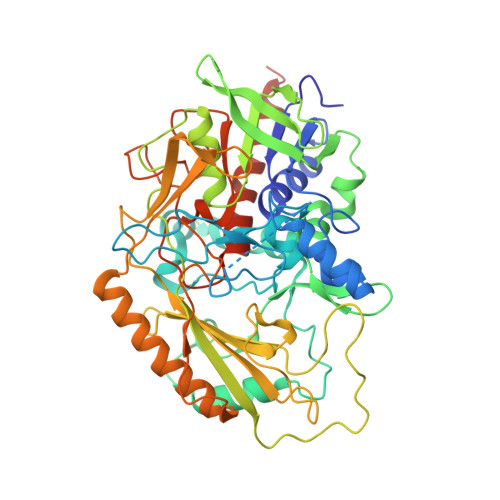
    C -glycosides have a unique structure, in which an anomeric carbon of a sugar is directly bonded to the carbon of an aglycone skeleton. One of the natural C -glycosides, carminic acid, is utilized by the food, cosmetic, and pharmaceutical industries, for a total of more than 200 tons/y worldwide. However, a metabolic pathway of carminic acid has never been identified. In this study, we isolated the previously unknown carminic acid-catabolizing microorganism and discovered a flavoenzyme " C -glycoside 3-oxidase" named CarA that catalyzes oxidation of the sugar moiety of carminic acid. A Basic Local Alignment Search Tool (BLAST) search demonstrated that CarA homologs were distributed in soil microorganisms but not intestinal ones. In addition to CarA, two CarA homologs were cloned and heterologously expressed, and their biochemical properties were determined. Furthermore, a crystal structure of one homolog was determined. Together with the biochemical analysis, the crystal structure and a mutagenesis analysis of CarA revealed the mechanisms underlying their substrate specificity and catalytic reaction. Our study suggests that CarA and its homologs play a crucial role in the metabolism of C -glycosides in nature.


  • Organizational Affiliation

    Graduate School of Life and Environmental Sciences, University of Tsukuba, Tsukuba, Ibaraki 305-8572, Japan.


Macromolecules
Find similar proteins by:  (by identity cutoff)  |  3D Structure
Entity ID: 1
MoleculeChains Sequence LengthOrganismDetailsImage
6'''-hydroxyparomomycin C oxidase
A, B
528Microbacterium trichothecenolyticumMutation(s): 0 
Gene Names: livQ_3RS82_01892
EC: 1.1.3
UniProt
Find proteins for A0A0M2HFA3 (Microbacterium trichothecenolyticum)
Explore A0A0M2HFA3 
Go to UniProtKB:  A0A0M2HFA3
Entity Groups  
Sequence Clusters30% Identity50% Identity70% Identity90% Identity95% Identity100% Identity
UniProt GroupA0A0M2HFA3
Sequence Annotations
Expand
  • Reference Sequence
Small Molecules
Experimental Data & Validation

Experimental Data

  • Method: X-RAY DIFFRACTION
  • Resolution: 2.40 Å
  • R-Value Free: 0.257 
  • R-Value Work: 0.224 
  • R-Value Observed: 0.226 
  • Space Group: H 3 2
Unit Cell:
Length ( Å )Angle ( ˚ )
a = 267.081α = 90
b = 267.081β = 90
c = 87.209γ = 120
Software Package:
Software NamePurpose
XDSdata reduction
XSCALEdata scaling
BALBESphasing
Cootmodel building
PHENIXrefinement

Structure Validation

View Full Validation Report


Ligand Structure Quality Assessment 


Entry History & Funding Information

Deposition Data


Funding OrganizationLocationGrant Number
Japan Agency for Medical Research and Development (AMED)JapanJP20am0101001

Revision History  (Full details and data files)

  • Version 1.0: 2021-09-08
    Type: Initial release
  • Version 1.1: 2022-03-02
    Changes: Database references
  • Version 1.2: 2023-11-29
    Changes: Data collection, Refinement description