7DMB

Crystal structure of trans-methyltransferase CalH complex with SAH


Experimental Data Snapshot

  • Method: X-RAY DIFFRACTION
  • Resolution: 2.10 Å
  • R-Value Free: 0.243 
  • R-Value Work: 0.186 
  • R-Value Observed: 0.189 

wwPDB Validation   3D Report Full Report


Ligand Structure Quality Assessment 


This is version 1.2 of the entry. See complete history


Literature

One Polyketide Synthase, Two Distinct Products: Trans-Acting Enzyme-Controlled Product Divergence in Calbistrin Biosynthesis.

Tao, H.Mori, T.Wei, X.Matsuda, Y.Abe, I.

(2021) Angew Chem Int Ed Engl 60: 8851-8858

  • DOI: https://doi.org/10.1002/anie.202016525
  • Primary Citation of Related Structures:  
    7DMB

  • PubMed Abstract: 

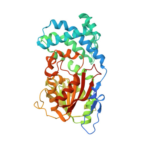
    Calbistrins are fungal polyketides consisting of the characteristic decalin and polyene moieties. Although the biosynthetic gene cluster of calbistrin A was recently identified, the pathway of calbistrin A biosynthesis has largely remained uninvestigated. Herein, we investigated the mechanism by which the backbone structures of calbistrins are formed, by heterologous and in vitro reconstitution of the biosynthesis and a structural biological study. Intriguingly, our analyses revealed that the decalin and polyene portions of calbistrins are synthesized by the single polyketide synthase (PKS) CalA, with the aid of the trans-acting enoylreductase CalK and the trans-acting C-methyltransferase CalH, respectively. We also determined that the esterification of the two polyketide parts is catalyzed by the acyltransferase CalD. Our study has uncovered a novel dual-functional PKS and thus broadened our understanding of how fungi synthesize diverse polyketide natural products.


  • Organizational Affiliation

    Graduate School of Pharmaceutical Sciences, The University of Tokyo, 7-3-1 Hongo, Bunkyo-ku, Tokyo, 113-0033, Japan.


Macromolecules
Find similar proteins by:  (by identity cutoff)  |  3D Structure
Entity ID: 1
MoleculeChains Sequence LengthOrganismDetailsImage
Putative methyltransferase
A, B
386Aspergillus stellatusMutation(s): 0 
UniProt
Find proteins for A0A679DLY7 (Emericella variicolor)
Explore A0A679DLY7 
Go to UniProtKB:  A0A679DLY7
Entity Groups  
Sequence Clusters30% Identity50% Identity70% Identity90% Identity95% Identity100% Identity
UniProt GroupA0A679DLY7
Sequence Annotations
Expand
  • Reference Sequence
Experimental Data & Validation

Experimental Data

  • Method: X-RAY DIFFRACTION
  • Resolution: 2.10 Å
  • R-Value Free: 0.243 
  • R-Value Work: 0.186 
  • R-Value Observed: 0.189 
  • Space Group: P 1
Unit Cell:
Length ( Å )Angle ( ˚ )
a = 41.81α = 87.786
b = 60.594β = 88.34
c = 71.146γ = 89.742
Software Package:
Software NamePurpose
XDSdata reduction
PHENIXrefinement
Aimlessdata scaling

Structure Validation

View Full Validation Report


Ligand Structure Quality Assessment 


Entry History 

Deposition Data

Revision History  (Full details and data files)

  • Version 1.0: 2021-02-10
    Type: Initial release
  • Version 1.1: 2021-04-14
    Changes: Database references
  • Version 1.2: 2024-03-27
    Changes: Data collection, Database references