7DLQ

CRYSTAL STRUCTURE OF THE COMPLEX OF LACTOPEROXIDASE WITH HYDROGEN PEROXIDE AT 1.77A RESOLUTION


Experimental Data Snapshot

  • Method: X-RAY DIFFRACTION
  • Resolution: 1.77 Å
  • R-Value Free: 0.226 
  • R-Value Work: 0.187 

wwPDB Validation   3D Report Full Report


Ligand Structure Quality Assessment 


This is version 1.3 of the entry. See complete history


Literature

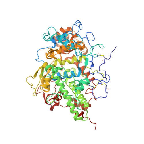
Structure of a ternary complex of lactoperoxidase with iodide and hydrogen peroxide at 1.77 angstrom resolution.

Singh, P.K.Sharma, P.Bhushan, A.Kaur, P.Sharma, S.Singh, T.P.

(2021) J Inorg Biochem 220: 111461-111461

  • DOI: https://doi.org/10.1016/j.jinorgbio.2021.111461
  • Primary Citation of Related Structures:  
    7DLQ, 7DN6, 7DN7

  • PubMed Abstract: 

    Lactoperoxidase (LPO) is a mammalian heme peroxidase which catalyzes the conversion of thiocyanate (SCN¯) and iodide (I - ) by hydrogen peroxide (H 2 O 2 ) into antimicrobial hypothiocyanite (OSCN¯) and hypoiodite (IO - ). The prosthetic heme group is covalently attached to LPO through two ester linkages involving conserved glutamate and aspartate residues. On the proximal side, His351 is coordinated to heme iron while His 109 is located in the substrate binding site on the distal heme side. We report here the first structure of the ternary complex of LPO with iodide (I - ) and H 2 O 2 at 1.77 Å resolution. LPO was crystallized with ammonium iodide and the crystals were soaked in the reservoir solution containing H 2 O 2. Structure determination showed the presence of an iodide ion and a H 2 O 2 molecule in the substrate binding site. The iodide ion occupied the position which is stabilized by the interactions with heme moiety, His109, Arg255 and Glu258 while H 2 O 2 was held between the heme iron and His109. The presence of I - in the distal heme cavity seems to screen the positive charge of Arg255 thus suppressing the proton transfer from H 2 O 2 to His109. This prevents compound I formation and allows trapping of a stable enzyme-substrate (LPO-I - -H 2 O 2) ternary complex. This stable geometrical arrangement of H 2 O 2 in the distal heme cavity of LPO is similar to that of H 2 O 2 in the structure of the transient intermediate of the palm tree heme peroxidase. The biochemical studies showed that the catalytic activity of LPO decreased when the samples of LPO were preincubated with ammonium iodide.


  • Organizational Affiliation

    Department of Biophysics, All India Institute of Medical Sciences, New Delhi, India.


Macromolecules
Find similar proteins by:  (by identity cutoff)  |  3D Structure
Entity ID: 1
MoleculeChains Sequence LengthOrganismDetailsImage
Lactoperoxidase595Bos taurusMutation(s): 0 
EC: 1.11.1.7
UniProt
Find proteins for P80025 (Bos taurus)
Explore P80025 
Go to UniProtKB:  P80025
Entity Groups  
Sequence Clusters30% Identity50% Identity70% Identity90% Identity95% Identity100% Identity
UniProt GroupP80025
Sequence Annotations
Expand
  • Reference Sequence
Oligosaccharides

Help

Entity ID: 2
MoleculeChains Length2D Diagram Glycosylation3D Interactions
2-acetamido-2-deoxy-beta-D-glucopyranose-(1-4)-2-acetamido-2-deoxy-beta-D-glucopyranose
B, C
2N-Glycosylation
Glycosylation Resources
GlyTouCan:  G42666HT
GlyCosmos:  G42666HT
GlyGen:  G42666HT
Small Molecules
Ligands 9 Unique
IDChains Name / Formula / InChI Key2D Diagram3D Interactions
HEM (Subject of Investigation/LOI)
Query on HEM

Download Ideal Coordinates CCD File 
D [auth A]PROTOPORPHYRIN IX CONTAINING FE
C34 H32 Fe N4 O4
KABFMIBPWCXCRK-RGGAHWMASA-L
NAG
Query on NAG

Download Ideal Coordinates CCD File 
F [auth A]2-acetamido-2-deoxy-beta-D-glucopyranose
C8 H15 N O6
OVRNDRQMDRJTHS-FMDGEEDCSA-N
IOD
Query on IOD

Download Ideal Coordinates CCD File 
AA [auth A]
BA [auth A]
CA [auth A]
EA [auth A]
FA [auth A]
AA [auth A],
BA [auth A],
CA [auth A],
EA [auth A],
FA [auth A],
G [auth A],
H [auth A],
I [auth A],
J [auth A],
K [auth A],
L [auth A],
M [auth A],
N [auth A],
O [auth A],
P [auth A],
W [auth A],
X [auth A],
Y [auth A],
Z [auth A]
IODIDE ION
I
XMBWDFGMSWQBCA-UHFFFAOYSA-M
GOL
Query on GOL

Download Ideal Coordinates CCD File 
Q [auth A]GLYCEROL
C3 H8 O3
PEDCQBHIVMGVHV-UHFFFAOYSA-N
OSM
Query on OSM

Download Ideal Coordinates CCD File 
T [auth A],
U [auth A],
V [auth A]
1-(OXIDOSULFANYL)METHANAMINE
C H5 N O S
KONFWJXIYYGXOO-UHFFFAOYSA-N
ZN
Query on ZN

Download Ideal Coordinates CCD File 
S [auth A]ZINC ION
Zn
PTFCDOFLOPIGGS-UHFFFAOYSA-N
EDO
Query on EDO

Download Ideal Coordinates CCD File 
R [auth A]1,2-ETHANEDIOL
C2 H6 O2
LYCAIKOWRPUZTN-UHFFFAOYSA-N
CA
Query on CA

Download Ideal Coordinates CCD File 
E [auth A]CALCIUM ION
Ca
BHPQYMZQTOCNFJ-UHFFFAOYSA-N
PEO
Query on PEO

Download Ideal Coordinates CCD File 
DA [auth A]HYDROGEN PEROXIDE
H2 O2
MHAJPDPJQMAIIY-UHFFFAOYSA-N
Experimental Data & Validation

Experimental Data

  • Method: X-RAY DIFFRACTION
  • Resolution: 1.77 Å
  • R-Value Free: 0.226 
  • R-Value Work: 0.187 
  • Space Group: P 1 21 1
Unit Cell:
Length ( Å )Angle ( ˚ )
a = 54.231α = 90
b = 79.837β = 102.67
c = 77.548γ = 90
Software Package:
Software NamePurpose
REFMACrefinement
HKL-2000data reduction
SCALEPACKdata scaling
MOLREPphasing

Structure Validation

View Full Validation Report


Ligand Structure Quality Assessment 


Entry History 

Deposition Data

Revision History  (Full details and data files)

  • Version 1.0: 2020-12-16
    Type: Initial release
  • Version 1.1: 2021-04-28
    Changes: Database references
  • Version 1.2: 2021-05-05
    Changes: Database references
  • Version 1.3: 2023-11-29
    Changes: Data collection, Database references, Derived calculations, Refinement description