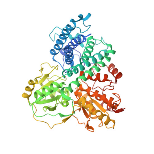
Crystal structure of Escherichia coli class II hybrid cluster protein, HCP, reveals a [4Fe-4S] cluster at the N-terminal protrusion.
Fujishiro, T., Ooi, M., Takaoka, K.(2021) FEBS J 288: 6752-6768
- PubMed: 34101368 Search on PubMed
- DOI: https://doi.org/10.1111/febs.16062
- Primary Citation Related Structures:
7DE4 - PubMed Abstract:
Hybrid cluster protein (HCP) is a unique Fe-S-O-type metallocluster-containing enzyme present in many anaerobic organisms and is categorized into three distinct classes (I, II, and III). The class II HCP uniquely utilizes hybrid cluster protein reductase (HCR), unlike the other classes of HCPs. To gain structural insights into the electron transfer system between the class II HCP and HCR, we elucidated the X-ray crystal structure of Escherichia coli HCP (Ec HCP), representing the first report of a class II HCP structure. Surprisingly, Ec HCP was found to harbor a [4Fe-4S] cluster rather than a [2Fe-2S] cluster at the N-terminal Cys-rich region, similar to class I HCPs. It was also found that the Cys-rich motif forms a unique protrusion and that the surrounding charge distributions on the surface of class II Ec HCP are distinct from those of class I HCPs. The functional significance of the Cys-rich region was investigated using an Ec HCP variant (chimeric HCP) containing a class I HCP Cys-rich motif from Desulfovibrio desulfuricans. The biochemical analyses showed that the chimeric HCP lacks the hybrid cluster and the electron-accepting function from HCR despite the formation of the chimeric HCP-HCR complex. Furthermore, HCP-HCR molecular docking analysis suggested that the protrusion area serves as an HCR-binding region. Therefore, the protrusion of the unique Cys-rich motif and the surrounding area of class II HCP are likely important for maturation of Ec HCP and orienting HCR onto the surface of HCP to facilitate electron transfer in the HCP-HCR complex.
- Department of Biochemistry and Molecular Biology, Graduate School of Science and Engineering, Saitama University, Saitama, Japan.
Organizational Affiliation: