7DB6 | pdb_00007db6

human melatonin receptor MT1 - Gi1 complex


Experimental Data Snapshot

  • Method: ELECTRON MICROSCOPY
  • Resolution: 3.30 Å
  • Aggregation State: PARTICLE 
  • Reconstruction Method: SINGLE PARTICLE 

wwPDB Validation 3D Report Full Report

Validation slider image for 7DB6

This is version 1.3 of the entry. See complete history. 

Literature

Cryo-EM structure of the human MT 1 -G i signaling complex.

Okamoto, H.H., Miyauchi, H., Inoue, A., Raimondi, F., Tsujimoto, H., Kusakizako, T., Shihoya, W., Yamashita, K., Suno, R., Nomura, N., Kobayashi, T., Iwata, S., Nishizawa, T., Nureki, O.

(2021) Nat Struct Mol Biol 28: 694-701

  • DOI: https://doi.org/10.1038/s41594-021-00634-1
  • Primary Citation Related Structures: 
    7DB6

  • PubMed Abstract: 

    Melatonin receptors (MT 1 and MT 2 ) transduce inhibitory signaling by melatonin (N-acetyl-5-methoxytryptamine), which is associated with sleep induction and circadian rhythm modulation. Although recently reported crystal structures of ligand-bound MT 1 and MT 2 elucidated the basis of ligand entry and recognition, the ligand-induced MT 1 rearrangement leading to G i -coupling remains unclear. Here we report a cryo-EM structure of the human MT 1 -G i signaling complex at 3.3 Å resolution, revealing melatonin-induced conformational changes propagated to the G-protein-coupling interface during activation. In contrast to other G i -coupled receptors, MT 1 exhibits a large outward movement of TM6, which is considered a specific feature of G s -coupled receptors. Structural comparison of G i and G s complexes demonstrated conformational diversity of the C-terminal entry of the G i protein, suggesting loose and variable interactions at the end of the α5 helix of G i protein. These notions, together with our biochemical and computational analyses, highlight variable binding modes of Gα i and provide the basis for the selectivity of G-protein signaling.


  • Organizational Affiliation: 
    • Department of Biological Sciences, Graduate School of Science, The University of Tokyo, Bunkyo-ku, Tokyo, Japan.

Macromolecule Content 

  • Total Structure Weight: 155.96 kDa 
  • Atom Count: 8,767 
  • Modeled Residue Count: 1,116 
  • Deposited Residue Count: 1,398 
  • Unique protein chains: 5

Macromolecules

Find similar proteins by:|  3D Structure
Entity ID: 1
MoleculeChains  Sequence LengthOrganismDetailsImage
Guanine nucleotide-binding protein G(i) subunit alpha-1354Homo sapiensMutation(s): 0 
Gene Names: GNAI1
EC: 3.6.5
Membrane Entity: Yes 
UniProt & NIH Common Fund Data Resources
Find proteins for P63096 (Homo sapiens)
Explore P63096 
Go to UniProtKB:  P63096
PHAROS:  P63096
GTEx:  ENSG00000127955 
Entity Groups
Sequence Clusters30% Identity50% Identity70% Identity90% Identity95% Identity100% Identity
UniProt GroupP63096
Sequence Annotations
Expand
Reference Sequence
Find similar proteins by:|  3D Structure
Entity ID: 2
MoleculeChains  Sequence LengthOrganismDetailsImage
Guanine nucleotide-binding protein G(I)/G(S)/G(T) subunit beta-1344Rattus rattusMutation(s): 0 
UniProt & NIH Common Fund Data Resources
Find proteins for P62873 (Homo sapiens)
Explore P62873 
Go to UniProtKB:  P62873
PHAROS:  P62873
GTEx:  ENSG00000078369 
Entity Groups
Sequence Clusters30% Identity50% Identity70% Identity90% Identity95% Identity100% Identity
UniProt GroupP62873
Sequence Annotations
Expand
Reference Sequence
Find similar proteins by:|  3D Structure
Entity ID: 3
MoleculeChains  Sequence LengthOrganismDetailsImage
Guanine nucleotide-binding protein G(I)/G(S)/G(O) subunit gamma-268Bos taurusMutation(s): 0 
Gene Names: GNG2
Membrane Entity: Yes 
UniProt
Find proteins for P63212 (Bos taurus)
Explore P63212 
Go to UniProtKB:  P63212
Entity Groups
Sequence Clusters30% Identity50% Identity70% Identity90% Identity95% Identity100% Identity
UniProt GroupP63212
Sequence Annotations
Expand
Reference Sequence
Find similar proteins by:|  3D Structure
Entity ID: 4
MoleculeChains  Sequence LengthOrganismDetailsImage
scFv16D [auth E]260Mus musculusMutation(s): 0 
Entity Groups
Sequence Clusters30% Identity50% Identity70% Identity90% Identity95% Identity100% Identity
Sequence Annotations
Expand
Reference Sequence
Find similar proteins by:|  3D Structure
Entity ID: 5
MoleculeChains  Sequence LengthOrganismDetailsImage
Melatonin receptor type 1AE [auth D]372Homo sapiensMutation(s): 0 
Gene Names: MTNR1A
Membrane Entity: Yes 
UniProt & NIH Common Fund Data Resources
Find proteins for P48039 (Homo sapiens)
Explore P48039 
Go to UniProtKB:  P48039
PHAROS:  P48039
GTEx:  ENSG00000168412 
Entity Groups
Sequence Clusters30% Identity50% Identity70% Identity90% Identity95% Identity100% Identity
UniProt GroupP48039
Sequence Annotations
Expand
Reference Sequence

Small Molecules

Ligands 1 Unique
IDChains Name / Formula / InChI Key2D Diagram3D Interactions
JEV
(Subject of Investigation/LOI)

Query on JEV


Download:Ideal Coordinates CCD File
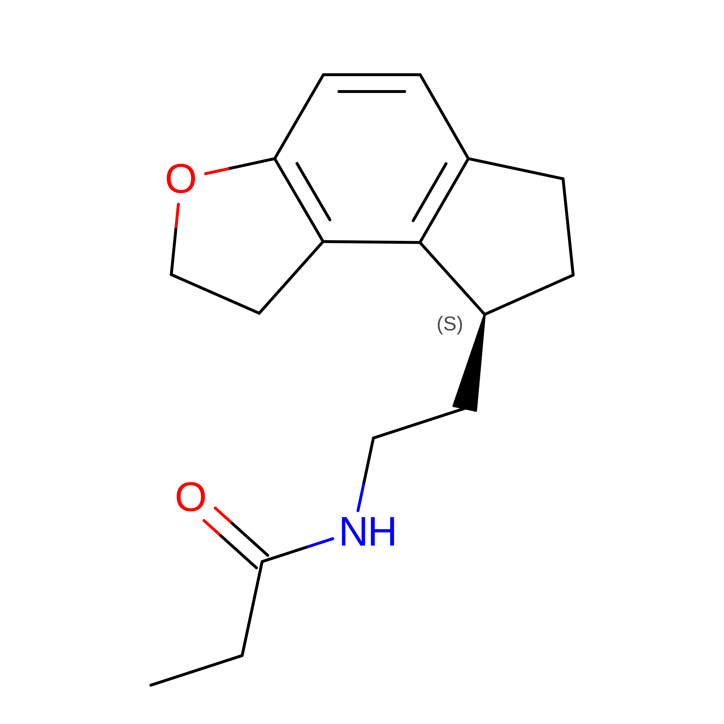
F [auth D]N-{2-[(8S)-1,6,7,8-tetrahydro-2H-indeno[5,4-b]furan-8-yl]ethyl}propanamide
C16 H21 N O2
YLXDSYKOBKBWJQ-LBPRGKRZSA-N
Binding Affinity Annotations 
IDSourceBinding Affinity
JEV BindingDB:  7DB6 Ki: min: 0.01, max: 0.01 (nM) from 2 assay(s)

Experimental Data & Validation

Experimental Data

  • Method: ELECTRON MICROSCOPY
  • Resolution: 3.30 Å
  • Aggregation State: PARTICLE 
  • Reconstruction Method: SINGLE PARTICLE 
EM Software:
TaskSoftware PackageVersion
MODEL REFINEMENTPHENIX

Structure Validation

View Full Validation Report


Entry History 

Deposition Data

Revision History  (Full details and data files)

  • Version 1.0: 2021-08-18
    Type: Initial release
  • Version 1.1: 2022-02-16
    Changes: Database references
  • Version 1.2: 2024-11-13
    Changes: Data collection, Structure summary
  • Version 1.3: 2025-07-02
    Changes: Data collection