7D9N

Crystal structure of a non-canonic progeria mutation S143F at lamin A/C and its structural implication to the premature aging


Experimental Data Snapshot

  • Method: X-RAY DIFFRACTION
  • Resolution: 3.70 Å
  • R-Value Free: 0.304 
  • R-Value Work: 0.287 
  • R-Value Observed: 0.289 

wwPDB Validation   3D Report Full Report


This is version 1.2 of the entry. See complete history


Literature

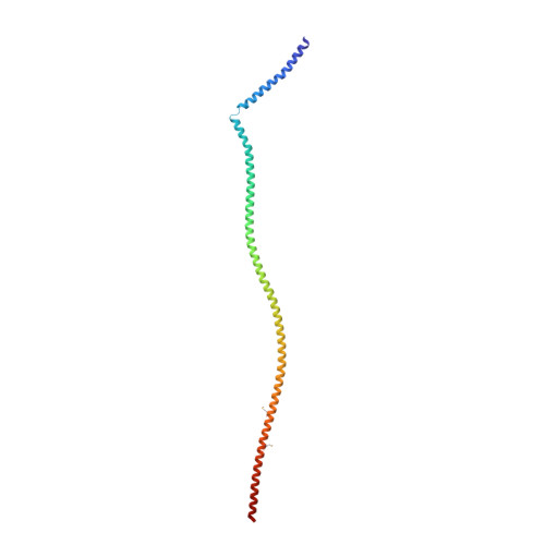
Crystal structure of progeria mutant S143F lamin A/C reveals increased hydrophobicity driving nuclear deformation.

Ahn, J.Jeong, S.Kang, S.M.Jo, I.Park, B.J.Ha, N.C.

(2022) Commun Biol 5: 267-267

  • DOI: https://doi.org/10.1038/s42003-022-03212-3
  • Primary Citation of Related Structures:  
    7D9N

  • PubMed Abstract: 

    Lamins are intermediate filaments that form a 3-D meshwork in the periphery of the nuclear envelope. The recent crystal structure of a long fragment of human lamin A/C visualized the tetrameric assembly unit of the central rod domain as a polymerization intermediate. A genetic mutation of S143F caused a phenotype characterized by both progeria and muscular dystrophy. In this study, we determined the crystal structure of the lamin A/C fragment harboring the S143F mutation. The obtained structure revealed the X-shaped interaction between the tetrameric units in the crystals, potentiated by the hydrophobic interactions of the mutated Phe143 residues. Subsequent studies indicated that the X-shaped interaction between the filaments plays a crucial role in disrupting the normal lamin meshwork. Our findings suggest the assembly mechanism of the 3-D meshwork and further provide a molecular framework for understanding the aging process by nuclear deformation.


  • Organizational Affiliation

    Department of Agricultural Biotechnology, Center for Food and Bioconvergence, and Research Institute of Agriculture and Life Sciences, CALS, Seoul National University, Seoul, 08826, Republic of Korea.


Macromolecules
Find similar proteins by:  (by identity cutoff)  |  3D Structure
Entity ID: 1
MoleculeChains Sequence LengthOrganismDetailsImage
Lamin-A/C
A, B
204Homo sapiensMutation(s): 1 
Gene Names: LMNALMN1
UniProt & NIH Common Fund Data Resources
Find proteins for P02545 (Homo sapiens)
Explore P02545 
Go to UniProtKB:  P02545
PHAROS:  P02545
GTEx:  ENSG00000160789 
Entity Groups  
Sequence Clusters30% Identity50% Identity70% Identity90% Identity95% Identity100% Identity
UniProt GroupP02545
Sequence Annotations
Expand
  • Reference Sequence
Small Molecules
Modified Residues  1 Unique
IDChains TypeFormula2D DiagramParent
MSE
Query on MSE
A, B
L-PEPTIDE LINKINGC5 H11 N O2 SeMET
Experimental Data & Validation

Experimental Data

  • Method: X-RAY DIFFRACTION
  • Resolution: 3.70 Å
  • R-Value Free: 0.304 
  • R-Value Work: 0.287 
  • R-Value Observed: 0.289 
  • Space Group: I 41 2 2
Unit Cell:
Length ( Å )Angle ( ˚ )
a = 123.808α = 90
b = 123.808β = 90
c = 458.545γ = 90
Software Package:
Software NamePurpose
PHENIXrefinement
DENZOdata reduction
HKL-2000data scaling
PHENIXphasing

Structure Validation

View Full Validation Report


Entry History & Funding Information

Deposition Data


Funding OrganizationLocationGrant Number
National Research Foundation (NRF, Korea)Korea, Republic Of2019R1A2C208513512
National Research Foundation (NRF, Korea)Korea, Republic Of2020R1A4A101932211

Revision History  (Full details and data files)

  • Version 1.0: 2021-11-17
    Type: Initial release
  • Version 1.1: 2022-11-30
    Changes: Database references
  • Version 1.2: 2023-11-29
    Changes: Data collection, Refinement description