7D6A

Crystal structure of Oryza sativa Os4BGlu18 monolignol beta-glucosidase


Experimental Data Snapshot

  • Method: X-RAY DIFFRACTION
  • Resolution: 1.70 Å
  • R-Value Free: 0.175 
  • R-Value Work: 0.150 
  • R-Value Observed: 0.151 

wwPDB Validation   3D Report Full Report


Ligand Structure Quality Assessment 


This is version 1.2 of the entry. See complete history


Literature

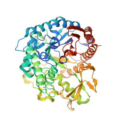
Structural analysis of rice Os4BGlu18 monolignol beta-glucosidase.

Baiya, S.Pengthaisong, S.Kitjaruwankul, S.Ketudat Cairns, J.R.

(2021) PLoS One 16: e0241325-e0241325

  • DOI: https://doi.org/10.1371/journal.pone.0241325
  • Primary Citation of Related Structures:  
    7D6A, 7D6B

  • PubMed Abstract: 

    Monolignol glucosides are storage forms of monolignols, which are polymerized to lignin to strengthen plant cell walls. The conversion of monolignol glucosides to monolignols is catalyzed by monolignol β-glucosidases. Rice Os4BGlu18 β-glucosidase catalyzes hydrolysis of the monolignol glucosides, coniferin, syringin, and p-coumaryl alcohol glucoside more efficiently than other natural substrates. To understand more clearly the basis for substrate specificity of a monolignol β-glucosidase, the structure of Os4BGlu18 was determined by X-ray crystallography. Crystals of Os4BGlu18 and its complex with δ-gluconolactone diffracted to 1.7 and 2.1 Å resolution, respectively. Two protein molecules were found in the asymmetric unit of the P212121 space group of their isomorphous crystals. The Os4BGlu18 structure exhibited the typical (β/α)8 TIM barrel of glycoside hydrolase family 1 (GH1), but the four variable loops and two disulfide bonds appeared significantly different from other known structures of GH1 β-glucosidases. Molecular docking studies of the Os4BGlu18 structure with monolignol substrate ligands placed the glycone in a similar position to the δ-gluconolactone in the complex structure and revealed the interactions between protein and ligands. Molecular docking, multiple sequence alignment, and homology modeling identified amino acid residues at the aglycone-binding site involved in substrate specificity for monolignol β-glucosides. Thus, the structural basis of substrate recognition and hydrolysis by monolignol β-glucosidases was elucidated.


  • Organizational Affiliation

    Faculty of Science at Sriracha, Kasetsart University, Sriracha Campus, Sriracha, Chonburi, Thailand.


Macromolecules
Find similar proteins by:  (by identity cutoff)  |  3D Structure
Entity ID: 1
MoleculeChains Sequence LengthOrganismDetailsImage
Beta-glucosidase 18
A, B
482Oryza sativa Japonica GroupMutation(s): 0 
Gene Names: BGLU18Os04g0513900LOC_Os04g43410OSJNBa0004N05.26OSJNBb0070J16.3
EC: 3.2.1.21
UniProt
Find proteins for Q7XSK0 (Oryza sativa subsp. japonica)
Explore Q7XSK0 
Go to UniProtKB:  Q7XSK0
Entity Groups  
Sequence Clusters30% Identity50% Identity70% Identity90% Identity95% Identity100% Identity
UniProt GroupQ7XSK0
Sequence Annotations
Expand
  • Reference Sequence
Small Molecules
Ligands 3 Unique
IDChains Name / Formula / InChI Key2D Diagram3D Interactions
MES (Subject of Investigation/LOI)
Query on MES

Download Ideal Coordinates CCD File 
C [auth A],
M [auth B]
2-(N-MORPHOLINO)-ETHANESULFONIC ACID
C6 H13 N O4 S
SXGZJKUKBWWHRA-UHFFFAOYSA-N
GOL (Subject of Investigation/LOI)
Query on GOL

Download Ideal Coordinates CCD File 
D [auth A]
E [auth A]
F [auth A]
G [auth A]
H [auth A]
D [auth A],
E [auth A],
F [auth A],
G [auth A],
H [auth A],
I [auth A],
J [auth A],
K [auth A],
N [auth B],
O [auth B],
P [auth B],
Q [auth B],
R [auth B]
GLYCEROL
C3 H8 O3
PEDCQBHIVMGVHV-UHFFFAOYSA-N
ZN (Subject of Investigation/LOI)
Query on ZN

Download Ideal Coordinates CCD File 
L [auth B]ZINC ION
Zn
PTFCDOFLOPIGGS-UHFFFAOYSA-N
Experimental Data & Validation

Experimental Data

  • Method: X-RAY DIFFRACTION
  • Resolution: 1.70 Å
  • R-Value Free: 0.175 
  • R-Value Work: 0.150 
  • R-Value Observed: 0.151 
  • Space Group: P 21 21 21
Unit Cell:
Length ( Å )Angle ( ˚ )
a = 52.134α = 90
b = 83.759β = 90
c = 207.649γ = 90
Software Package:
Software NamePurpose
REFMACrefinement
HKL-3000data reduction
HKL-3000data scaling
MOLREPphasing

Structure Validation

View Full Validation Report


Ligand Structure Quality Assessment 


Entry History & Funding Information

Deposition Data


Funding OrganizationLocationGrant Number
Other governmentThailandBRG5980015 Thailand Research Fund and Suranaree University of Technology
Other governmentThailandSuranaree University of Technology

Revision History  (Full details and data files)

  • Version 1.0: 2021-01-13
    Type: Initial release
  • Version 1.1: 2021-02-10
    Changes: Database references
  • Version 1.2: 2023-11-29
    Changes: Data collection, Database references, Refinement description