7D4B | pdb_00007d4b

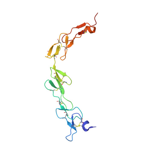
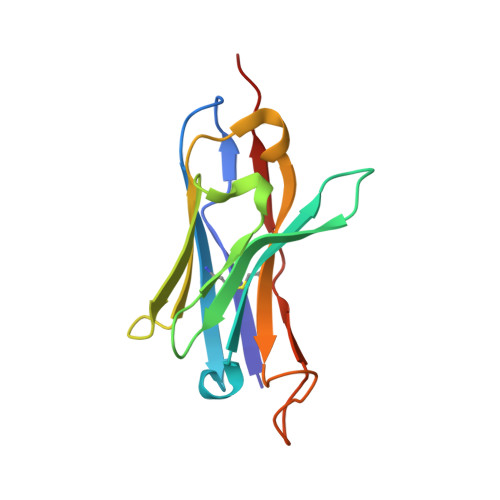
Crystal structure of 4-1BB in complex with a VHH


Experimental Data Snapshot

  • Method: X-RAY DIFFRACTION
  • Resolution: 3.14 Å
  • R-Value Free: 
    0.258 (Depositor), 0.257 (DCC) 
  • R-Value Work: 
    0.211 (Depositor), 0.219 (DCC) 
  • R-Value Observed: 
    0.214 (Depositor) 

Starting Models: experimental
View more details

wwPDB Validation 3D Report Full Report

Validation slider image for 7D4B

This is version 1.2 of the entry. See complete history. 

Literature

Generation of a safe and efficacious llama single-domain antibody fragment (vHH) targeting the membrane-proximal region of 4-1BB for engineering therapeutic bispecific antibodies for cancer.

Zhai, T., Wang, C., Xu, Y., Huang, W., Yuan, Z., Wang, T., Dai, S., Peng, S., Pang, T., Jiang, W., Huang, Y., Zou, Y., Xu, Y., Sun, J., Gong, X., Zhang, J., Tsun, A., Li, B., Miao, X.

(2021) J Immunother Cancer 9

  • DOI: https://doi.org/10.1136/jitc-2020-002131
  • Primary Citation Related Structures: 
    7CZD, 7D4B

  • PubMed Abstract: 

    The discovery of checkpoint inhibitors towards cytotoxic T-lymphocyte protein 4 (CTLA-4) and programmed cell death protein 1 (PD-1) has been revolutionary for the treatment of cancers. These therapies have only offered an average of 20%-30% response rates across the tumor spectrum and the combination of agonists towards the tumor-necrosis superfamily members, such as 4-1BB and CD40, has shown potent efficacy in preclinical studies; however, these agonists have exhibited high degrees of toxicity with limited efficacy in human trials. In this study, we have generated a single-domain antibody towards a unique epitope of 4-1BB that limits its potential on-target toxicity while maintaining sufficient potency. This 4-1BB binder is ideal for use in the engineering of multispecific antibodies to localize 4-1BB activation within the tumor microenvironment, as shown here by a anti-PD-L1/4-1BB bispecific candidate (PM1003). To determine the functional activity of the 4-1BB- and PD-L1-binding elements of PM1003, in vitro luciferase reporter and primary cell assays were used to test the potency of programmed cell death 1 ligand 1 (PD-L1) blockade and PD-L1-mediated 4-1BB activation via cross-bridging. X-ray crystallography was conducted to resolve the binding epitopes of the respective binding arms, and accurate binding kinetics were determined using standard affinity measurement techniques. Human 4-1BB and/or PD-L1 knock-in mice were used in cancer models for testing the in vivo antitumor efficacy of PM1003, and safety was evaluated further. PM1003 shows potent activation of 4-1BB and blockade of PD-L1 in cell-based assays. 4-1BB activation was exerted through the bridging of PD-L1 on target cells and 4-1BB on effector cells. No PD-L1-independent activation of 4-1BB was observed. Through X-ray crystallography, a unique binding epitope in the cysteine-rich domain 4 (CRD4) region was resolved that provides high potency and potentially low on-target toxicity as determined by primary immune cell assays and toxicity evaluation in vivo . A unique single-domain antibody was discovered that binds to the CRD4 domain of 4-1BB. When incorporated into a 4-1BB/PD-L1 bispecific (PM1003), we have shown the potent inhibition of PD-L1 activity with 4-1BB agonism upon cross-bridging with PD-L1 in vitro . Antitumor activity with minimal toxicity was found in vivo . Thus, PM1003 is a uniquely differentiating and next generation therapeutic agent for cancer therapy.


  • Organizational Affiliation: 
    • Shanghai Institute of Immunology, Department of Immunology and Microbiology, Shanghai Jiao Tong University School of Medicine, Shanghai, China.

Macromolecule Content 

  • Total Structure Weight: 31.21 kDa 
  • Atom Count: 2,182 
  • Modeled Residue Count: 265 
  • Deposited Residue Count: 267 
  • Unique protein chains: 2

Macromolecules

Find similar proteins by:|  3D Structure
Entity ID: 1
MoleculeChains  Sequence LengthOrganismDetailsImage
Tumor necrosis factor receptor superfamily member 9140Homo sapiensMutation(s): 0 
Gene Names: TNFRSF9, CD137, ILA
UniProt & NIH Common Fund Data Resources
Find proteins for Q07011 (Homo sapiens)
Explore Q07011 
Go to UniProtKB:  Q07011
GTEx:  ENSG00000049249 
Entity Groups
Sequence Clusters30% Identity50% Identity70% Identity90% Identity95% Identity100% Identity
UniProt GroupQ07011
Glycosylation
Glycosylation Sites: 2Go to GlyGen: Q07011-1
Sequence Annotations
Expand
Reference Sequence
Find similar proteins by:|  3D Structure
Entity ID: 2
MoleculeChains  Sequence LengthOrganismDetailsImage
anti-4-1BB VHH127Lama glamaMutation(s): 0 
Entity Groups
Sequence Clusters30% Identity50% Identity70% Identity90% Identity95% Identity100% Identity
Sequence Annotations
Expand
Reference Sequence

Oligosaccharides

Help  
Entity ID: 3
MoleculeChains Length2D Diagram GlycosylationD Interactions
alpha-D-mannopyranose-(1-3)-alpha-D-mannopyranose-(1-6)-[alpha-D-mannopyranose-(1-3)]beta-D-mannopyranose-(1-4)-2-acetamido-2-deoxy-beta-D-glucopyranose-(1-4)-[alpha-L-fucopyranose-(1-6)]2-acetamido-2-deoxy-beta-D-glucopyranose
C
7N-Glycosylation
Glycosylation Resources
GlyTouCan: G32156ZV
GlyCosmos: G32156ZV
GlyGen: G32156ZV
Entity ID: 4
MoleculeChains Length2D Diagram GlycosylationD Interactions
2-acetamido-2-deoxy-beta-D-glucopyranose-(1-4)-[alpha-L-fucopyranose-(1-6)]2-acetamido-2-deoxy-beta-D-glucopyranose
D
3N-Glycosylation
Glycosylation Resources
GlyTouCan: G21290RB
GlyCosmos: G21290RB
GlyGen: G21290RB

Small Molecules

Ligands 3 Unique
IDChains Name / Formula / InChI Key2D Diagram3D Interactions
SO4

Query on SO4


Download:Ideal Coordinates CCD File
G [auth A],
H [auth A],
L [auth B]
SULFATE ION
O4 S
QAOWNCQODCNURD-UHFFFAOYSA-L
EDO

Query on EDO


Download:Ideal Coordinates CCD File
J [auth B],
K [auth B]
1,2-ETHANEDIOL
C2 H6 O2
LYCAIKOWRPUZTN-UHFFFAOYSA-N
CL

Query on CL


Download:Ideal Coordinates CCD File
E [auth A],
F [auth A],
I [auth B]
CHLORIDE ION
Cl
VEXZGXHMUGYJMC-UHFFFAOYSA-M

Experimental Data & Validation

Experimental Data

  • Method: X-RAY DIFFRACTION
  • Resolution: 3.14 Å
  • R-Value Free:  0.258 (Depositor), 0.257 (DCC) 
  • R-Value Work:  0.211 (Depositor), 0.219 (DCC) 
  • R-Value Observed: 0.214 (Depositor) 
Space Group: P 41 21 2
Unit Cell:
Length ( Å )Angle ( ˚ )
a = 106.851α = 90
b = 106.851β = 90
c = 146.41γ = 90
Software Package:
Software NamePurpose
REFMACrefinement
PDB_EXTRACTdata extraction
Aimlessdata scaling
PHASERphasing

Structure Validation

View Full Validation Report


Entry History 

Deposition Data

  • Released Date: 2021-07-14 
  • Deposition Author(s): Wang, C.

Revision History  (Full details and data files)

  • Version 1.0: 2021-07-14
    Type: Initial release
  • Version 1.1: 2023-11-29
    Changes: Data collection, Database references, Refinement description
  • Version 1.2: 2024-10-16
    Changes: Structure summary