7CY8

Crystal Structure of CMD1 in complex with 5mC-DNA and vitamin C


Experimental Data Snapshot

  • Method: X-RAY DIFFRACTION
  • Resolution: 2.40 Å
  • R-Value Free: 0.235 
  • R-Value Work: 0.190 
  • R-Value Observed: 0.192 

wwPDB Validation   3D Report Full Report


Ligand Structure Quality Assessment 


This is version 1.3 of the entry. See complete history


Literature

Molecular mechanism for vitamin C-derived C 5 -glyceryl-methylcytosine DNA modification catalyzed by algal TET homologue CMD1.

Li, W.Zhang, T.Sun, M.Shi, Y.Zhang, X.J.Xu, G.L.Ding, J.

(2021) Nat Commun 12: 744-744

  • DOI: https://doi.org/10.1038/s41467-021-21061-2
  • Primary Citation of Related Structures:  
    7CY4, 7CY5, 7CY6, 7CY7, 7CY8

  • PubMed Abstract: 

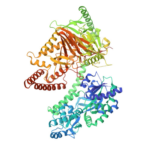
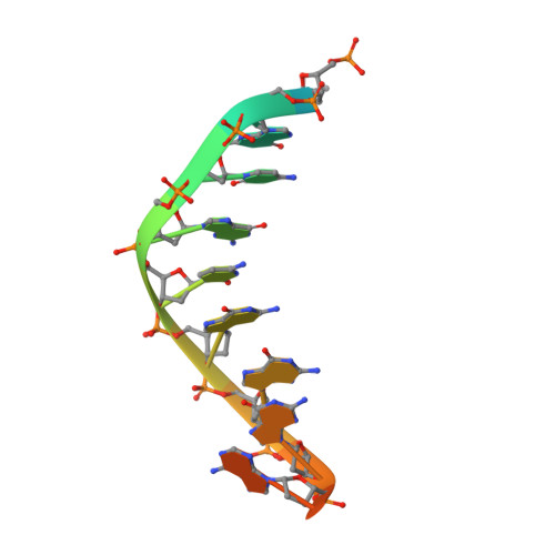
    C 5 -glyceryl-methylcytosine (5gmC) is a novel DNA modification catalyzed by algal TET homologue CMD1 using vitamin C (VC) as co-substrate. Here, we report the structures of CMD1 in apo form and in complexes with VC or/and dsDNA. CMD1 exhibits comparable binding affinities for DNAs of different lengths, structures, and 5mC levels, and displays a moderate substrate preference for 5mCpG-containing DNA. CMD1 adopts the typical DSBH fold of Fe 2+ /2-OG-dependent dioxygenases. The lactone form of VC binds to the active site and mono-coordinates the Fe 2+ in a manner different from 2-OG. The dsDNA binds to a positively charged cleft of CMD1 and the 5mC/C is inserted into the active site and recognized by CMD1 in a similar manner as the TET proteins. The functions of key residues are validated by mutagenesis and activity assay. Our structural and biochemical data together reveal the molecular mechanism for the VC-derived 5gmC DNA modification by CMD1.


  • Organizational Affiliation

    State Key Laboratory of Molecular Biology, Shanghai Institute of Biochemistry and Cell Biology, Center for Excellence in Molecular Cell Science, Chinese Academy of Sciences, University of Chinese Academy of Sciences, Shanghai, China.


Macromolecules

Find similar proteins by:  (by identity cutoff)  |  3D Structure
Entity ID: 1
MoleculeChains Sequence LengthOrganismDetailsImage
Maltodextrin-binding protein,5-methylcytosine-modifying enzyme 1917Escherichia coliChlamydomonas reinhardtii
This entity is chimeric
Mutation(s): 7 
Gene Names: 
EC: 1.14.99
UniProt
Find proteins for P0AEX9 (Escherichia coli (strain K12))
Explore P0AEX9 
Go to UniProtKB:  P0AEX9
Find proteins for A0A2K3D5Z7 (Chlamydomonas reinhardtii)
Explore A0A2K3D5Z7 
Go to UniProtKB:  A0A2K3D5Z7
Entity Groups  
Sequence Clusters30% Identity50% Identity70% Identity90% Identity95% Identity100% Identity
UniProt GroupsA0A2K3D5Z7P0AEX9
Sequence Annotations
Expand
  • Reference Sequence

Find similar nucleic acids by:  Sequence   |   3D Structure  

Entity ID: 2
MoleculeChains LengthOrganismImage
DNA (5'-D(P*(5CM)P*GP*CP*GP*CP*GP*GP*GP*A)-3')B [auth C],
C [auth D]
14synthetic construct
Sequence Annotations
Expand
  • Reference Sequence
Experimental Data & Validation

Experimental Data

  • Method: X-RAY DIFFRACTION
  • Resolution: 2.40 Å
  • R-Value Free: 0.235 
  • R-Value Work: 0.190 
  • R-Value Observed: 0.192 
  • Space Group: C 1 2 1
Unit Cell:
Length ( Å )Angle ( ˚ )
a = 154.488α = 90
b = 125.646β = 102.96
c = 64.058γ = 90
Software Package:
Software NamePurpose
REFMACrefinement
XDSdata scaling
PDB_EXTRACTdata extraction
XDSdata reduction
PHENIXphasing

Structure Validation

View Full Validation Report


Ligand Structure Quality Assessment 


Entry History & Funding Information

Deposition Data


Funding OrganizationLocationGrant Number
National Natural Science Foundation of China (NSFC)China31530013
National Natural Science Foundation of China (NSFC)China31800622
Chinese Academy of SciencesChinaXDB37030305

Revision History  (Full details and data files)

  • Version 1.0: 2020-12-30
    Type: Initial release
  • Version 1.1: 2021-02-10
    Changes: Database references
  • Version 1.2: 2021-02-17
    Changes: Database references
  • Version 1.3: 2023-11-29
    Changes: Data collection, Database references, Refinement description