7CNS

Crystal structure of Thermococcus kodakaraensis aconitase X (holo-form)


Experimental Data Snapshot

  • Method: X-RAY DIFFRACTION
  • Resolution: 1.90 Å
  • R-Value Free: 0.201 
  • R-Value Work: 0.169 
  • R-Value Observed: 0.171 

wwPDB Validation   3D Report Full Report


Ligand Structure Quality Assessment 


This is version 1.1 of the entry. See complete history


Literature

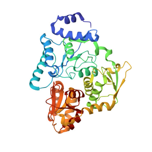
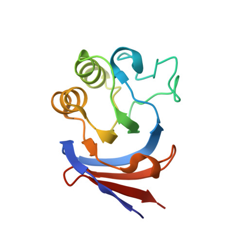
Crystal structures of aconitase X enzymes from bacteria and archaea provide insights into the molecular evolution of the aconitase superfamily.

Watanabe, S.Murase, Y.Watanabe, Y.Sakurai, Y.Tajima, K.

(2021) Commun Biol 4: 687-687

  • DOI: https://doi.org/10.1038/s42003-021-02147-5
  • Primary Citation of Related Structures:  
    7CNP, 7CNQ, 7CNR, 7CNS, 7D2R

  • PubMed Abstract: 

    Aconitase superfamily members catalyze the homologous isomerization of specific substrates by sequential dehydration and hydration and contain a [4Fe-4S] cluster. However, monomeric and heterodimeric types of function unknown aconitase X (AcnX) have recently been characterized as a cis-3-hydroxy-L-proline dehydratase (AcnX Type-I ) and mevalonate 5-phosphate dehydratase (AcnX Type-II ), respectively. We herein elucidated the crystal structures of AcnX Type-I from Agrobacterium tumefaciens (AtAcnX) and AcnX Type-II from Thermococcus kodakarensis (TkAcnX) without a ligand and in complex with substrates. AtAcnX and TkAcnX contained the [2Fe-2S] and [3Fe-4S] clusters, respectively, conforming to UV and EPR spectroscopy analyses. The binding sites of the [Fe-S] cluster and substrate were clearlydifferent from those that were completely conserved in other aconitase enzymes; however, theoverall structural frameworks and locations of active sites were partially similar to each other.These results provide novel insights into the evolutionary scenario of the aconitase superfamilybased on the recruitment hypothesis.


  • Organizational Affiliation

    Department of Bioscience, Graduate School of Agriculture, Ehime University, Matsuyama, Ehime, Japan. irab@agr.ehime-u.ac.jp.


Macromolecules
Find similar proteins by:  (by identity cutoff)  |  3D Structure
Entity ID: 1
MoleculeChains Sequence LengthOrganismDetailsImage
DUF521 domain-containing protein386Thermococcus kodakarensis KOD1Mutation(s): 0 
Gene Names: TK1249
UniProt
Find proteins for Q5JGJ6 (Thermococcus kodakarensis (strain ATCC BAA-918 / JCM 12380 / KOD1))
Explore Q5JGJ6 
Go to UniProtKB:  Q5JGJ6
Entity Groups  
Sequence Clusters30% Identity50% Identity70% Identity90% Identity95% Identity100% Identity
UniProt GroupQ5JGJ6
Sequence Annotations
Expand
  • Reference Sequence
Find similar proteins by:  (by identity cutoff)  |  3D Structure
Entity ID: 2
MoleculeChains Sequence LengthOrganismDetailsImage
UPF0107 protein TK1248134Thermococcus kodakarensis KOD1Mutation(s): 0 
Gene Names: TK1248
UniProt
Find proteins for Q5JGJ7 (Thermococcus kodakarensis (strain ATCC BAA-918 / JCM 12380 / KOD1))
Explore Q5JGJ7 
Go to UniProtKB:  Q5JGJ7
Entity Groups  
Sequence Clusters30% Identity50% Identity70% Identity90% Identity95% Identity100% Identity
UniProt GroupQ5JGJ7
Sequence Annotations
Expand
  • Reference Sequence
Small Molecules
Experimental Data & Validation

Experimental Data

  • Method: X-RAY DIFFRACTION
  • Resolution: 1.90 Å
  • R-Value Free: 0.201 
  • R-Value Work: 0.169 
  • R-Value Observed: 0.171 
  • Space Group: P 42 21 2
Unit Cell:
Length ( Å )Angle ( ˚ )
a = 102.444α = 90
b = 102.444β = 90
c = 115.849γ = 90
Software Package:
Software NamePurpose
XDSdata scaling
PHENIXrefinement
PDB_EXTRACTdata extraction
PHASERphasing

Structure Validation

View Full Validation Report


Ligand Structure Quality Assessment 


Entry History 

Deposition Data

Revision History  (Full details and data files)

  • Version 1.0: 2021-06-16
    Type: Initial release
  • Version 1.1: 2022-12-28
    Changes: Database references