7CJP

Crystal structure of metal-free state of glucose isomerase

  • Classification: ISOMERASE
  • Organism(s): Streptomyces rubiginosus
  • Mutation(s): No 

  • Deposited: 2020-07-12 Released: 2021-07-14 
  • Deposition Author(s): Nam, K.H.
  • Funding Organization(s): National Research Foundation (NRF, Korea)

Experimental Data Snapshot

  • Method: X-RAY DIFFRACTION
  • Resolution: 1.50 Å
  • R-Value Free: 0.166 
  • R-Value Work: 0.149 
  • R-Value Observed: 0.149 

wwPDB Validation   3D Report Full Report


This is version 1.2 of the entry. See complete history


Literature

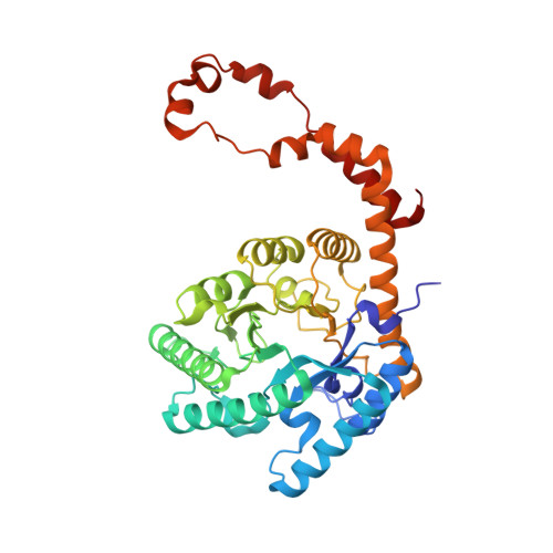
Crystal structure of the metal-free state of glucose isomerase reveals its minimal open configuration for metal binding.

Nam, K.H.

(2021) Biochem Biophys Res Commun 547: 69-74

  • DOI: https://doi.org/10.1016/j.bbrc.2021.02.026
  • Primary Citation of Related Structures:  
    7CJO, 7CJP

  • PubMed Abstract: 

    Glucose/xylose isomerase catalyzes the reversible isomerization of d-glucose and d-xylose to d-fructose and d-xylulose, respectively. This enzyme is not only involved in sugar metabolism but also has industrial applications, such as in the production of high fructose corn syrup and bioethanol. Various crystal structures of glucose isomerase have shown the binding configuration of the substrate and its molecular mechanism; however, the metal binding mechanism required for the isomerization reaction has not been fully elucidated. To better understand the functional metal binding, the crystal structures of the metal-bound and metal-free states of Streptomyces rubiginosus glucose isomerase (SruGI) were determined at 1.4 Å and 1.5 Å resolution, respectively. In the meal-bound state of SruGI, Mg 2+ is bound at the M1 and M2 sites, while in the metal-free state, these sites are occupied by water molecules. Structural comparison between the metal binding sites of the metal-bound and metal-free states of SruGI revealed that residues Glu217 and Asp257 exhibit a rigid configuration at the bottom of the metal binding site, suggesting that they serve as a metal-binding platform that defined the location of the metal. In contrast, the side chains of Glu218, His220, Asp255, Asp257, and Asp287 showed configuration changes such as shifts and rotations. Notably, in the metal-free state, the side chains of these amino acids are shifted away from the metal binding site, indicating that the metal-binding residues exhibit a minimal open configuration, which allows metal binding without large conformational changes.


  • Organizational Affiliation

    Department of Life Science, Pohang University of Science and Technology, Pohang, 37673, Republic of Korea. Electronic address: structures@postech.ac.kr.


Macromolecules
Find similar proteins by:  (by identity cutoff)  |  3D Structure
Entity ID: 1
MoleculeChains Sequence LengthOrganismDetailsImage
Xylose isomerase
A, B
388Streptomyces rubiginosusMutation(s): 0 
EC: 5.3.1.5
UniProt
Find proteins for P24300 (Streptomyces rubiginosus)
Explore P24300 
Go to UniProtKB:  P24300
Entity Groups  
Sequence Clusters30% Identity50% Identity70% Identity90% Identity95% Identity100% Identity
UniProt GroupP24300
Sequence Annotations
Expand
  • Reference Sequence
Small Molecules
Ligands 1 Unique
IDChains Name / Formula / InChI Key2D Diagram3D Interactions
EDO
Query on EDO

Download Ideal Coordinates CCD File 
C [auth A]
D [auth A]
E [auth A]
F [auth A]
G [auth A]
C [auth A],
D [auth A],
E [auth A],
F [auth A],
G [auth A],
H [auth A],
I [auth A],
J [auth A],
K [auth A],
L [auth A],
M [auth A],
N [auth A],
O [auth A],
P [auth B],
Q [auth B],
R [auth B],
S [auth B],
T [auth B],
U [auth B],
V [auth B],
W [auth B],
X [auth B],
Y [auth B]
1,2-ETHANEDIOL
C2 H6 O2
LYCAIKOWRPUZTN-UHFFFAOYSA-N
Experimental Data & Validation

Experimental Data

  • Method: X-RAY DIFFRACTION
  • Resolution: 1.50 Å
  • R-Value Free: 0.166 
  • R-Value Work: 0.149 
  • R-Value Observed: 0.149 
  • Space Group: P 21 21 2
Unit Cell:
Length ( Å )Angle ( ˚ )
a = 86.288α = 90
b = 93.009β = 90
c = 98.914γ = 90
Software Package:
Software NamePurpose
PHENIXrefinement
HKL-2000data scaling
PDB_EXTRACTdata extraction
HKL-2000data reduction
MOLREPphasing

Structure Validation

View Full Validation Report


Entry History & Funding Information

Deposition Data

  • Released Date: 2021-07-14 
  • Deposition Author(s): Nam, K.H.

Funding OrganizationLocationGrant Number
National Research Foundation (NRF, Korea)Korea, Republic OfNRF-2017R1D1A1B03033087
National Research Foundation (NRF, Korea)Korea, Republic OfNRF-2017M3A9F6029736

Revision History  (Full details and data files)

  • Version 1.0: 2021-07-14
    Type: Initial release
  • Version 1.1: 2023-01-25
    Changes: Database references
  • Version 1.2: 2023-11-29
    Changes: Data collection, Refinement description