Two Distinct Conformations in 34 FliF Subunits Generate Three Different Symmetries within the Flagellar MS-Ring.
Takekawa, N., Kawamoto, A., Sakuma, M., Kato, T., Kojima, S., Kinoshita, M., Minamino, T., Namba, K., Homma, M., Imada, K.(2021) mBio 12
- PubMed: 33653894 Search on PubMedSearch on PubMed Central
- DOI: https://doi.org/10.1128/mBio.03199-20
- Primary Citation Related Structures:
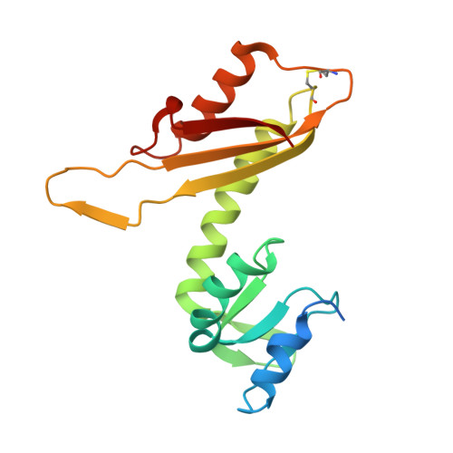
7CIK - PubMed Abstract:
The bacterial flagellum is a protein nanomachine essential for bacterial motility. The flagellar basal body contains several ring structures. The MS-ring is embedded in the cytoplasmic membrane and is formed at the earliest stage of flagellar formation to serve as the base for flagellar assembly as well as a housing for the flagellar protein export gate complex. The MS-ring is formed by FliF, which has two transmembrane helices and a large periplasmic region. A recent electron cryomicroscopy (cryoEM) study of the MS-ring formed by overexpressed FliF revealed a symmetry mismatch between the S-ring and inner part of the M-ring. However, the actual symmetry relation in the native MS-ring and positions of missing domains remain obscure. Here, we show the structure of the M-ring by combining cryoEM and X-ray crystallography. The crystal structure of the N-terminal half of the periplasmic region of FliF showed that it consists of two domains (D1 and D2) resembling PrgK D1/PrgH D2 and PrgK D2/PrgH D3 of the injectisome. CryoEM analysis revealed that the inner part of the M-ring shows a gear wheel-like density with the inner ring of C23 symmetry surrounded by cogs with C11 symmetry, to which 34 copies of FliF D1-D2 fitted well. We propose that FliF D1-D2 adopts two distinct orientations in the M-ring relative to the rest of FliF, with 23 chains forming the wheel and 11 chains forming the cogs, and the 34 chains come together to form the S-ring with C34 symmetry for multiple functions of the MS-ring. IMPORTANCE The bacterial flagellum is a motility organelle formed by tens of thousands of protein molecules. At the earliest stage of flagellar assembly, a transmembrane protein, FliF, forms the MS-ring in the cytoplasmic membrane as the base for flagellar assembly. Here, we solved the crystal structure of a FliF fragment. Electron cryomicroscopy (cryoEM) structural analysis of the MS-ring showed that the M-ring and S-ring have different rotational symmetries. By docking the crystal structure of the FliF fragment into the cryoEM density map of the entire MS-ring, we built a model of the whole periplasmic region of FliF and proposed that FliF adopts two distinct conformations to generate three distinct C11, C23, and C34 symmetries within the MS-ring for its multiple functions.
- Department of Macromolecular Science, Graduate School of Science, Osaka University, Toyonaka, Osaka, Japan.
Organizational Affiliation: