7CE5 | pdb_00007ce5

Methanol-PQQ bound methanol dehydrogenase (MDH) from Methylococcus capsulatus (Bath)


Experimental Data Snapshot

  • Method: X-RAY DIFFRACTION
  • Resolution: 1.80 Å
  • R-Value Free: 
    0.170 (Depositor), 0.172 (DCC) 
  • R-Value Work: 
    0.140 (Depositor) 

Starting Model: experimental
View more details

wwPDB Validation 3D Report Full Report

Validation slider image for 7CE5

Ligand Structure Quality Assessment 


This is version 1.2 of the entry. See complete history

Literature

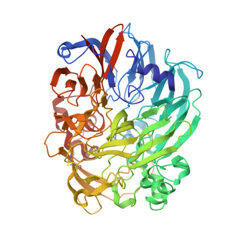
Mechanism of Pyrroloquinoline Quinone-Dependent Hydride Transfer Chemistry from Spectroscopic and High-Resolution X-ray Structural Studies of the Methanol Dehydrogenase from Methylococcus capsulatus (Bath).

Chan, S.I.Chuankhayan, P.Reddy Nareddy, P.K.Tsai, I.K.Tsai, Y.F.Chen, K.H.Yu, S.S.Chen, C.J.

(2021) J Am Chem Soc 143: 3359-3372

  • DOI: https://doi.org/10.1021/jacs.0c11414
  • Primary Citation Related Structures: 
    7CDL, 7CE5, 7CE9, 7CED, 7CFX

  • PubMed Abstract: 

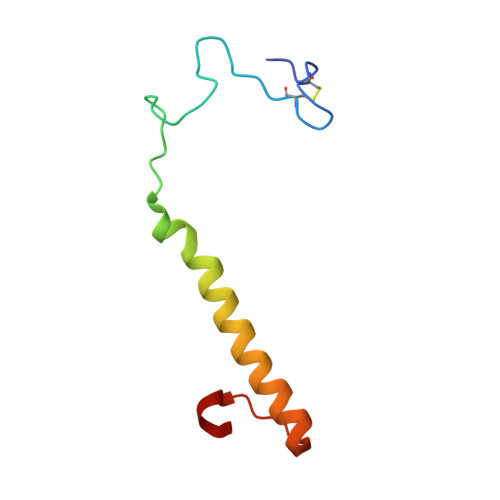
    The active site of methanol dehydrogenase (MDH) contains a rare disulfide bridge between adjacent cysteine residues. As a vicinal disulfide, the structure is highly strained, suggesting it might work together with the pyrroloquinoline quinone (PQQ) prosthetic group and the Ca 2+ ion in the catalytic turnover during methanol (CH 3 OH) oxidation. We purify MDH from Methylococcus capsulatus (Bath) with the disulfide bridge broken into two thiols. Spectroscopic and high-resolution X-ray crystallographic studies of this form of MDH indicate that the disulfide bridge is redox active. We observe an internal redox process within the holo -MDH that produces a disulfide radical anion concomitant with a companion PQQ radical, as evidenced by an optical absorption at 408 nm and a magnetically dipolar-coupled biradical in the EPR spectrum. These observations are corroborated by electron-density changes between the two cysteine sulfurs of the disulfide bridge as well as between the bound Ca 2+ ion and the O5-C5 bond of the PQQ in the high-resolution X-ray structure. On the basis of these findings, we propose a mechanism for the controlled redistribution of the two electrons during hydride transfer from the CH 3 OH in the alcohol oxidation without formation of the reduced PQQ ethenediol, a biradical mechanism that allows for possible recovery of the hydride for transfer to an external NAD + oxidant in the regeneration of the PQQ cofactor for multiple catalytic turnovers. In support of this mechanism, a steady-state level of the disulfide radical anion is observed during turnover of the MDH in the presence of CH 3 OH and NAD + .


  • Organizational Affiliation
    • Institute of Chemistry, Academia Sinica, Nangang, Taipei 11529, Taiwan.

Macromolecule Content 

  • Total Structure Weight: 578.56 kDa 
  • Atom Count: 46,034 
  • Modeled Residue Count: 5,152 
  • Deposited Residue Count: 5,160 
  • Unique protein chains: 2

Macromolecules

Find similar proteins by:|  3D Structure
Entity ID: 1
MoleculeChains  Sequence LengthOrganismDetailsImage
Methanol dehydrogenase protein, large subunit
A, B, C, D, G
A, B, C, D, G, H, M, N
573Methylococcus capsulatus str. BathMutation(s): 0 
UniProt
Find proteins for Q60AR6 (Methylococcus capsulatus (strain ATCC 33009 / NCIMB 11132 / Bath))
Explore Q60AR6 
Go to UniProtKB:  Q60AR6
Entity Groups
Sequence Clusters30% Identity50% Identity70% Identity90% Identity95% Identity100% Identity
UniProt GroupQ60AR6
Sequence Annotations
Expand
Reference Sequence
Find similar proteins by:|  3D Structure
Entity ID: 2
MoleculeChains  Sequence LengthOrganismDetailsImage
Methanol dehydrogenase [cytochrome c] subunit 2
E, F, I, J, K
E, F, I, J, K, L, O, P
72Methylococcus capsulatus str. BathMutation(s): 0 
EC: 1.1.2.7
UniProt
Find proteins for Q60AR3 (Methylococcus capsulatus (strain ATCC 33009 / NCIMB 11132 / Bath))
Explore Q60AR3 
Go to UniProtKB:  Q60AR3
Entity Groups
Sequence Clusters30% Identity50% Identity70% Identity90% Identity95% Identity100% Identity
UniProt GroupQ60AR3
Sequence Annotations
Expand
Reference Sequence

Small Molecules

Ligands 3 Unique
IDChains Name / Formula / InChI Key2D Diagram3D Interactions
PQQ
(Subject of Investigation/LOI)

Query on PQQ


Download:Ideal Coordinates CCD File
BA [auth D]
EA [auth G]
HA [auth H]
KA [auth M]
NA [auth N]
BA [auth D],
EA [auth G],
HA [auth H],
KA [auth M],
NA [auth N],
S [auth A],
V [auth B],
X [auth C]
PYRROLOQUINOLINE QUINONE
C14 H6 N2 O8
MMXZSJMASHPLLR-UHFFFAOYSA-N
CA

Query on CA


Download:Ideal Coordinates CCD File
CA [auth G]
FA [auth H]
IA [auth M]
LA [auth N]
Q [auth A]
CA [auth G],
FA [auth H],
IA [auth M],
LA [auth N],
Q [auth A],
T [auth B],
W [auth C],
Z [auth D]
CALCIUM ION
Ca
BHPQYMZQTOCNFJ-UHFFFAOYSA-N
MOH
(Subject of Investigation/LOI)

Query on MOH


Download:Ideal Coordinates CCD File
AA [auth D]
DA [auth G]
GA [auth H]
JA [auth M]
MA [auth N]
AA [auth D],
DA [auth G],
GA [auth H],
JA [auth M],
MA [auth N],
R [auth A],
U [auth B],
Y [auth C]
METHANOL
C H4 O
OKKJLVBELUTLKV-UHFFFAOYSA-N

Experimental Data & Validation

Experimental Data

  • Method: X-RAY DIFFRACTION
  • Resolution: 1.80 Å
  • R-Value Free:  0.170 (Depositor), 0.172 (DCC) 
  • R-Value Work:  0.140 (Depositor) 
Space Group: P 21 21 21
Unit Cell:
Length ( Å )Angle ( ˚ )
a = 128.612α = 90
b = 211.828β = 90
c = 223.912γ = 90
Software Package:
Software NamePurpose
REFMACrefinement
XDSdata reduction
XSCALEdata scaling
PHASERphasing

Structure Validation

View Full Validation Report


Ligand Structure Quality Assessment 


Entry History 

& Funding Information

Deposition Data


Funding OrganizationLocationGrant Number
Academia Sinica (Taiwan)Taiwan--

Revision History  (Full details and data files)

  • Version 1.0: 2021-06-30
    Type: Initial release
  • Version 1.1: 2023-11-29
    Changes: Data collection, Database references, Refinement description
  • Version 1.2: 2024-10-23
    Changes: Structure summary