


Structure of of a FIC-domain protein from Mycobacterium marinum
Kumar, S., Singh, A., Penmatsa, A., Surolia, A.To be published.
Experimental Data Snapshot
Starting Model: experimental
View more details
Entity ID: 1 | |||||
|---|---|---|---|---|---|
| Molecule | Chains | Sequence Length | Organism | Details | Image |
| Fic-domain containing protein | 412 | Mycobacterium marinum M | Mutation(s): 0 Gene Names: MMAR_0586 | 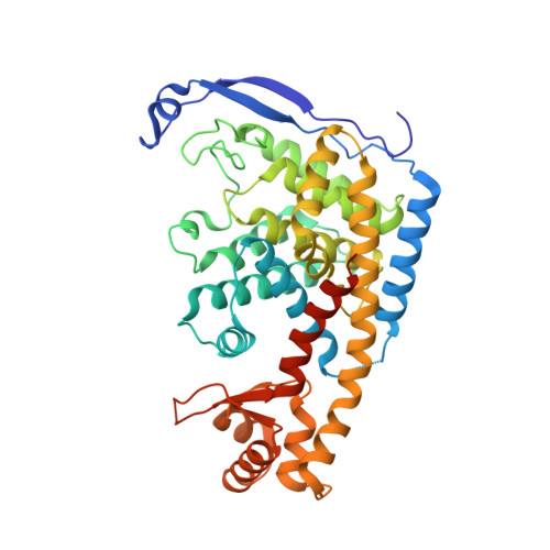 | |
UniProt | |||||
Entity Groups | |||||
| Sequence Clusters | 30% Identity50% Identity70% Identity90% Identity95% Identity100% Identity | ||||
| UniProt Group | B2HP08 | ||||
Sequence AnnotationsExpand | |||||
Reference Sequence | |||||
| Ligands 4 Unique | |||||
|---|---|---|---|---|---|
| ID | Chains | Name / Formula / InChI Key | 2D Diagram | 3D Interactions | |
| CDP (Subject of Investigation/LOI) Download:Ideal Coordinates CCD File | C [auth A], L [auth B] | CYTIDINE-5'-DIPHOSPHATE C9 H15 N3 O11 P2 ZWIADYZPOWUWEW-XVFCMESISA-N |  | ||
| SO4 Download:Ideal Coordinates CCD File | H [auth A], R [auth B], S [auth B] | SULFATE ION O4 S QAOWNCQODCNURD-UHFFFAOYSA-L |  | ||
| EDO Download:Ideal Coordinates CCD File | D [auth A] E [auth A] F [auth A] G [auth A] M [auth B] | 1,2-ETHANEDIOL C2 H6 O2 LYCAIKOWRPUZTN-UHFFFAOYSA-N |  | ||
| ACT Download:Ideal Coordinates CCD File | I [auth A], J [auth A], K [auth A], T [auth B], U [auth B] | ACETATE ION C2 H3 O2 QTBSBXVTEAMEQO-UHFFFAOYSA-M |  | ||
| Length ( Å ) | Angle ( ˚ ) |
|---|---|
| a = 76.066 | α = 90 |
| b = 107.9 | β = 90 |
| c = 110.588 | γ = 90 |
| Software Name | Purpose |
|---|---|
| PHENIX | refinement |
| XDS | data reduction |
| Aimless | data scaling |
| PHASER | phasing |
| Funding Organization | Location | Grant Number |
|---|---|---|
| Department of Biotechnology (DBT, India) | India | BT/PR5012/MED/29/419/2012 |
| Department of Biotechnology (DBT, India) | India | BT/PR24443/MED/2a/1220/2017 |


