7C72 | pdb_00007c72

Structure of a mycobacterium tuberculosis puromycin-hydrolyzing peptidase


Experimental Data Snapshot

  • Method: X-RAY DIFFRACTION
  • Resolution: 3.00 Å
  • R-Value Free: 
    0.218 (Depositor), 0.217 (DCC) 
  • R-Value Work: 
    0.177 (Depositor), 0.181 (DCC) 
  • R-Value Observed: 
    0.179 (Depositor) 

Starting Model: experimental
View more details

wwPDB Validation 3D Report Full Report

Validation slider image for 7C72

This is version 1.2 of the entry. See complete history

Literature

Mycobacterium tuberculosis puromycin hydrolase displays a prolyl oligopeptidase fold and an acyl aminopeptidase activity.

Zhao, Y.Feng, Q.Zhou, X.Zhang, Y.Lukman, M.Jiang, J.Ruiz-Carrillo, D.

(2021) Proteins 89: 614-622

  • DOI: https://doi.org/10.1002/prot.26044
  • Primary Citation Related Structures: 
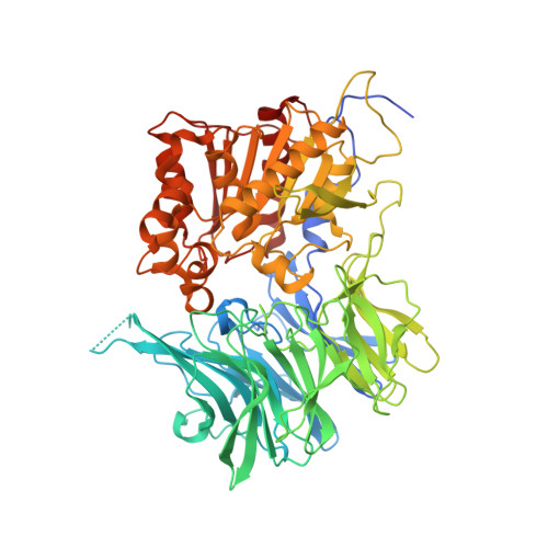
    7C72

  • PubMed Abstract: 

    Puromycin-hydrolizing peptidases have been described as members of the prolyl oligopeptidase peptidase family. These enzymes are present across all domains of life but still little is known of the homologs found in the pathogenic bacterium Mycobacterium tuberculosis. The crystal structure of a M. tuberculosis puromycin hydrolase peptidase has been determined at 3 Angstrom resolution, revealing a conserved prolyl oligopeptidase fold, defined by α/β-hydrolase and β-propeller domains with two distinctive loops that occlude access of large substrates to the active site. The enzyme displayed amino peptidase activity with a substrate specificity preference for hydrophobic residues in the decreasing order of phenylalanine, leucine, alanine and proline. The enzyme's active site is lined by residues Glu564 for the coordination of the substrates amino terminal moiety and His561, Val608, Tyr78, Trp306, Phe563 and Ty567 for the accommodation of hydrophobic substrates. The availability of a crystal structure for puromycin hydrolase of M. tuberculosis shall facilitate the development of inhibitors with therapeutic applications.


  • Organizational Affiliation
    • Department of Biological Sciences, School of Science, Xi'an Jiaotong-Liverpool University, Suzhou, Jiangsu, China.

Macromolecule Content 

  • Total Structure Weight: 151.31 kDa 
  • Atom Count: 10,076 
  • Modeled Residue Count: 1,302 
  • Deposited Residue Count: 1,392 
  • Unique protein chains: 1

Macromolecules

Find similar proteins by:|  3D Structure
Entity ID: 1
MoleculeChains  Sequence LengthOrganismDetailsImage
Prolyl oligopeptidase
A, B
696Mycobacterium tuberculosisMutation(s): 0 
Gene Names: CNE19725.1

Experimental Data & Validation

Experimental Data

  • Method: X-RAY DIFFRACTION
  • Resolution: 3.00 Å
  • R-Value Free:  0.218 (Depositor), 0.217 (DCC) 
  • R-Value Work:  0.177 (Depositor), 0.181 (DCC) 
  • R-Value Observed: 0.179 (Depositor) 
Space Group: P 63
Unit Cell:
Length ( Å )Angle ( ˚ )
a = 183.95α = 90
b = 183.95β = 90
c = 146.31γ = 120
Software Package:
Software NamePurpose
PHENIXrefinement
XDSdata reduction
XSCALEdata scaling
BALBESphasing

Structure Validation

View Full Validation Report


Entry History 

& Funding Information

Deposition Data


Funding OrganizationLocationGrant Number
Other privateChina--

Revision History  (Full details and data files)

  • Version 1.0: 2021-03-24
    Type: Initial release
  • Version 1.1: 2021-05-19
    Changes: Database references
  • Version 1.2: 2023-11-29
    Changes: Data collection, Database references, Refinement description