7BCW

Structure of MsbA in Salipro with ADP vanadate


Experimental Data Snapshot

  • Method: ELECTRON MICROSCOPY
  • Resolution: 3.50 Å
  • Aggregation State: PARTICLE 
  • Reconstruction Method: SINGLE PARTICLE 

wwPDB Validation   3D Report Full Report


This is version 1.2 of the entry. See complete history


Literature

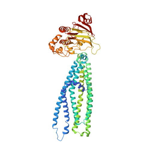
Cryo-EM structure of MsbA in saposin-lipid nanoparticles (Salipro) provides insights into nucleotide coordination.

Kehlenbeck, D.M.Traore, D.A.K.Josts, I.Sander, S.Moulin, M.Haertlein, M.Prevost, S.Forsyth, V.T.Tidow, H.

(2022) FEBS J 289: 2959-2970

  • DOI: https://doi.org/10.1111/febs.16327
  • Primary Citation of Related Structures:  
    7BCW

  • PubMed Abstract: 

    The ATP-binding cassette transporter MsbA is a lipid flippase, translocating lipid A, glycolipids, and lipopolysaccharides from the inner to the outer leaflet of the inner membrane of Gram-negative bacteria. It has been used as a model system for time-resolved structural studies as several MsbA structures in different states and reconstitution systems (detergent/nanodiscs/peptidiscs) are available. However, due to the limited resolution of the available structures, detailed structural information on the bound nucleotides has remained elusive. Here, we have reconstituted MsbA in saposin A-lipoprotein nanoparticles (Salipro) and determined the structure of ADP-vanadate-bound MsbA by single-particle cryo-electron microscopy to 3.5 Å resolution. This procedure has resulted in significantly improved resolution and enabled us to model all side chains and visualise detailed ADP-vanadate interactions in the nucleotide-binding domains. The approach may be applicable to other dynamic membrane proteins.


  • Organizational Affiliation

    The Hamburg Advanced Research Center for Bioorganic Chemistry (HARBOR), Germany.


Macromolecules
Find similar proteins by:  (by identity cutoff)  |  3D Structure
Entity ID: 1
MoleculeChains Sequence LengthOrganismDetailsImage
ATP-dependent lipid A-core flippase
A, B
598Escherichia coli K-12Mutation(s): 0 
Gene Names: msbAb0914JW0897
EC: 7.5.2.6
Membrane Entity: Yes 
UniProt
Find proteins for P60752 (Escherichia coli (strain K12))
Explore P60752 
Go to UniProtKB:  P60752
Entity Groups  
Sequence Clusters30% Identity50% Identity70% Identity90% Identity95% Identity100% Identity
UniProt GroupP60752
Sequence Annotations
Expand
  • Reference Sequence
Small Molecules
Ligands 4 Unique
IDChains Name / Formula / InChI Key2D Diagram3D Interactions
POV
Query on POV

Download Ideal Coordinates CCD File 
F [auth A],
J [auth B]
(2S)-3-(hexadecanoyloxy)-2-[(9Z)-octadec-9-enoyloxy]propyl 2-(trimethylammonio)ethyl phosphate
C42 H82 N O8 P
WTJKGGKOPKCXLL-PFDVCBLKSA-N
ADP (Subject of Investigation/LOI)
Query on ADP

Download Ideal Coordinates CCD File 
D [auth A],
H [auth B]
ADENOSINE-5'-DIPHOSPHATE
C10 H15 N5 O10 P2
XTWYTFMLZFPYCI-KQYNXXCUSA-N
VO4 (Subject of Investigation/LOI)
Query on VO4

Download Ideal Coordinates CCD File 
C [auth A],
G [auth B]
VANADATE ION
O4 V
LSGOVYNHVSXFFJ-UHFFFAOYSA-N
MG
Query on MG

Download Ideal Coordinates CCD File 
E [auth A],
I [auth B]
MAGNESIUM ION
Mg
JLVVSXFLKOJNIY-UHFFFAOYSA-N
Experimental Data & Validation

Experimental Data

  • Method: ELECTRON MICROSCOPY
  • Resolution: 3.50 Å
  • Aggregation State: PARTICLE 
  • Reconstruction Method: SINGLE PARTICLE 
EM Software:
TaskSoftware PackageVersion
RECONSTRUCTIONcryoSPARC2.15

Structure Validation

View Full Validation Report


Entry History 

Deposition Data

Revision History  (Full details and data files)

  • Version 1.0: 2022-01-12
    Type: Initial release
  • Version 1.1: 2022-01-19
    Changes: Database references
  • Version 1.2: 2022-05-25
    Changes: Database references