7BC4

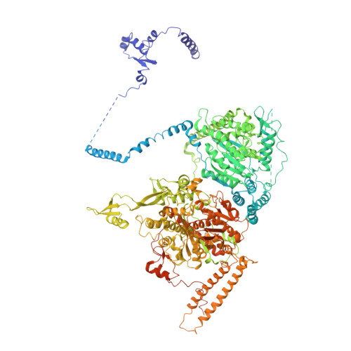
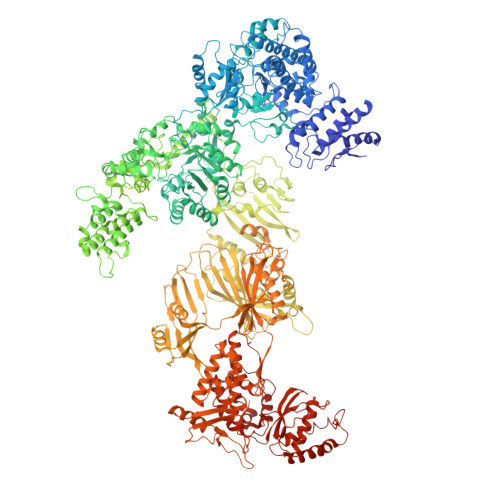
Cryo-EM structure of fatty acid synthase (FAS) from Pichia pastoris


Experimental Data Snapshot

  • Method: ELECTRON MICROSCOPY
  • Resolution: 3.10 Å
  • Aggregation State: PARTICLE 
  • Reconstruction Method: SINGLE PARTICLE 

wwPDB Validation   3D Report Full Report


This is version 1.0 of the entry. See complete history


Literature

Structural insight into Pichia pastoris fatty acid synthase.

Snowden, J.S.Alzahrani, J.Sherry, L.Stacey, M.Rowlands, D.J.Ranson, N.A.Stonehouse, N.J.

(2021) Sci Rep 11: 9773-9773

  • DOI: https://doi.org/10.1038/s41598-021-89196-2
  • Primary Citation of Related Structures:  
    7BC4, 7BC5

  • PubMed Abstract: 

    Type I fatty acid synthases (FASs) are critical metabolic enzymes which are common targets for bioengineering in the production of biofuels and other products. Serendipitously, we identified FAS as a contaminant in a cryoEM dataset of virus-like particles (VLPs) purified from P. pastoris, an important model organism and common expression system used in protein production. From these data, we determined the structure of P. pastoris FAS to 3.1 Å resolution. While the overall organisation of the complex was typical of type I FASs, we identified several differences in both structural and enzymatic domains through comparison with the prototypical yeast FAS from S. cerevisiae. Using focussed classification, we were also able to resolve and model the mobile acyl-carrier protein (ACP) domain, which is key for function. Ultimately, the structure reported here will be a useful resource for further efforts to engineer yeast FAS for synthesis of alternate products.


  • Organizational Affiliation

    Astbury Centre for Structural Molecular Biology, School of Molecular and Cellular Biology, Faculty of Biological Sciences, University of Leeds, Leeds, LS2 9JT, UK.


Macromolecules
Find similar proteins by:  (by identity cutoff)  |  3D Structure
Entity ID: 1
MoleculeChains Sequence LengthOrganismDetailsImage
Fatty acid synthase subunit alpha1,879Komagataella phaffii GS115Mutation(s): 0 
EC: 2.3.1.86 (PDB Primary Data), 1.1.1.100 (PDB Primary Data), 2.3.1.41 (PDB Primary Data)
UniProt
Find proteins for C4QY10 (Komagataella phaffii (strain GS115 / ATCC 20864))
Explore C4QY10 
Go to UniProtKB:  C4QY10
Entity Groups  
Sequence Clusters30% Identity50% Identity70% Identity90% Identity95% Identity100% Identity
UniProt GroupC4QY10
Sequence Annotations
Expand
  • Reference Sequence
Find similar proteins by:  (by identity cutoff)  |  3D Structure
Entity ID: 2
MoleculeChains Sequence LengthOrganismDetailsImage
Fatty acid synthase subunit beta2,069Komagataella phaffii GS115Mutation(s): 0 
EC: 2.3.1.86 (PDB Primary Data), 4.2.1.59 (PDB Primary Data), 1.3.1.9 (PDB Primary Data), 2.3.1.38 (PDB Primary Data), 2.3.1.39 (PDB Primary Data), 3.1.2.14 (PDB Primary Data)
UniProt
Find proteins for C4QVT8 (Komagataella phaffii (strain GS115 / ATCC 20864))
Explore C4QVT8 
Go to UniProtKB:  C4QVT8
Entity Groups  
Sequence Clusters30% Identity50% Identity70% Identity90% Identity95% Identity100% Identity
UniProt GroupC4QVT8
Sequence Annotations
Expand
  • Reference Sequence
Small Molecules
Ligands 1 Unique
IDChains Name / Formula / InChI Key2D Diagram3D Interactions
FMN
Query on FMN

Download Ideal Coordinates CCD File 
C [auth B]FLAVIN MONONUCLEOTIDE
C17 H21 N4 O9 P
FVTCRASFADXXNN-SCRDCRAPSA-N
Experimental Data & Validation

Experimental Data

  • Method: ELECTRON MICROSCOPY
  • Resolution: 3.10 Å
  • Aggregation State: PARTICLE 
  • Reconstruction Method: SINGLE PARTICLE 
EM Software:
TaskSoftware PackageVersion
RECONSTRUCTIONRELION3.0.7
MODEL REFINEMENTPHENIX1.14

Structure Validation

View Full Validation Report


Entry History & Funding Information

Deposition Data


Funding OrganizationLocationGrant Number
Wellcome TrustUnited Kingdom102174/B/13/Z
World Health Organization (WHO)United Kingdom2019/883397-O
Saudi Ministry of EducationSaudi Arabia--

Revision History  (Full details and data files)

  • Version 1.0: 2021-05-19
    Type: Initial release