7AT1

CRYSTAL STRUCTURES OF ASPARTATE CARBAMOYLTRANSFERASE LIGATED WITH PHOSPHONOACETAMIDE, MALONATE, AND CTP OR ATP AT 2.8-ANGSTROMS RESOLUTION AND NEUTRAL P*H


Experimental Data Snapshot

  • Method: X-RAY DIFFRACTION
  • Resolution: 2.80 Å
  • R-Value Work: 0.183 

wwPDB Validation   3D Report Full Report


This is version 2.1 of the entry. See complete history


Literature

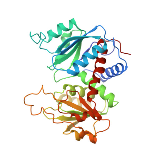
Crystal structures of aspartate carbamoyltransferase ligated with phosphonoacetamide, malonate, and CTP or ATP at 2.8-A resolution and neutral pH.

Gouaux, J.E.Stevens, R.C.Lipscomb, W.N.

(1990) Biochemistry 29: 7702-7715

  • DOI: https://doi.org/10.1021/bi00485a020
  • Primary Citation of Related Structures:  
    7AT1, 8AT1

  • PubMed Abstract: 

    The R-state structures of the ATP and CTP complexes of aspartate carbamoyltransferase ligated with phosphonoacetamide and malonate have been determined at 2.8-A resolution and neutral pH. These structures were solved by the method of molecular replacement and were refined to crystallographic residuals between 0.167 and 0.182. The triphosphate, the ribose, and the purine and pyrimidine moieties of ATP and CTP interact with similar regions of the allosteric domain of the regulatory dimer. ATP and CTP relatively increase and decrease the size of the allosteric site in the vicinity of the base, respectively. For both CTP and ATP at pH 7, the gamma-phosphates are bound to His20 and are also near Lys94, while the alpha-phosphates interact exclusively with Lys94. The 2'-hydroxyls of both CTP and ATP are near the amino group of Lys60. The pyrimidine ring of CTP makes specific hydrogen bonds at the allosteric site: the NH2 group donates hydrogen bonds to the main-chain carbonyls of Ile12 and Tyr89 and the pyrimidine ring carbonyl oxygen accepts a hydrogen bond from the amino group of Lys60; the nitrogen at position 3 in the pyrimidine ring is hydrogen bonded to a main-chain NH group of Ile12. The purine ring of ATP also makes numerous interactions with residues at the allosteric site: the purine NH2 (analogous to the amino group of CTP) donates a hydrogen bond to the main-chain carbonyl oxygen of Ile12, the N3 nitrogen interacts with the amino group of Lys60, and the N1 nitrogen hydrogen bonds to the NH group of Ile12. The binding of CTP and ATP to the allosteric site in the presence of phosphonoacetamide and malonate does not dramatically alter the structure of the allosteric binding site or of the allosteric domain. Nonetheless, in the CTP-ligated structure, the average separation between the catalytic trimers decreases by approximately 0.5 A, indicating a small shift of the quaternary structure toward the T state. In the CTP- and ATP-ligated R-state structures, the binding and occupancy of phosphonoacetamide and malonate are similar and the structures of the active sites are similar at the current resolution of 2.8 A.


  • Organizational Affiliation

    Gibbs Chemical Laboratory, Harvard University, Cambridge, Massachusetts 02138.


Macromolecules
Find similar proteins by:  (by identity cutoff)  |  3D Structure
Entity ID: 1
MoleculeChains Sequence LengthOrganismDetailsImage
ASPARTATE CARBAMOYLTRANSFERASE (R STATE), CATALYTIC CHAIN
A, C
310Escherichia coliMutation(s): 0 
EC: 2.1.3.2
UniProt
Find proteins for P0A786 (Escherichia coli (strain K12))
Explore P0A786 
Go to UniProtKB:  P0A786
Entity Groups  
Sequence Clusters30% Identity50% Identity70% Identity90% Identity95% Identity100% Identity
UniProt GroupP0A786
Sequence Annotations
Expand
  • Reference Sequence
Find similar proteins by:  (by identity cutoff)  |  3D Structure
Entity ID: 2
MoleculeChains Sequence LengthOrganismDetailsImage
ASPARTATE CARBAMOYLTRANSFERASE REGULATORY CHAIN
B, D
153Escherichia coliMutation(s): 0 
UniProt
Find proteins for P0A7F3 (Escherichia coli (strain K12))
Explore P0A7F3 
Go to UniProtKB:  P0A7F3
Entity Groups  
Sequence Clusters30% Identity50% Identity70% Identity90% Identity95% Identity100% Identity
UniProt GroupP0A7F3
Sequence Annotations
Expand
  • Reference Sequence
Oligosaccharides

Help

Entity ID: 3
MoleculeChains Length2D Diagram Glycosylation3D Interactions
alpha-D-glucopyranose-(1-4)-alpha-D-glucopyranose
E, F
2N/A
Glycosylation Resources
GlyTouCan:  G07411ON
GlyCosmos:  G07411ON
Small Molecules
Biologically Interesting Molecules (External Reference) 1 Unique
Experimental Data & Validation

Experimental Data

  • Method: X-RAY DIFFRACTION
  • Resolution: 2.80 Å
  • R-Value Work: 0.183 
  • Space Group: P 3 2 1
Unit Cell:
Length ( Å )Angle ( ˚ )
a = 122.5α = 90
b = 122.5β = 90
c = 156.5γ = 120
Software Package:
Software NamePurpose
X-PLORrefinement

Structure Validation

View Full Validation Report


Entry History 

Deposition Data

Revision History  (Full details and data files)

  • Version 1.0: 1990-10-15
    Type: Initial release
  • Version 1.1: 2008-03-03
    Changes: Version format compliance
  • Version 1.2: 2011-07-13
    Changes: Derived calculations, Version format compliance
  • Version 1.3: 2017-11-29
    Changes: Advisory, Derived calculations, Other
  • Version 2.0: 2020-07-29
    Type: Remediation
    Reason: Carbohydrate remediation
    Changes: Advisory, Atomic model, Data collection, Database references, Derived calculations, Non-polymer description, Structure summary
  • Version 2.1: 2024-03-06
    Changes: Data collection, Database references, Structure summary