7ALO | pdb_00007alo

Structure of B*27:09/photoRL9


Experimental Data Snapshot

  • Method: X-RAY DIFFRACTION
  • Resolution: 1.80 Å
  • R-Value Free: 
    0.266 (Depositor), 0.266 (DCC) 
  • R-Value Work: 
    0.223 (Depositor), 0.225 (DCC) 
  • R-Value Observed: 
    0.226 (Depositor) 

Starting Model: experimental
View more details

wwPDB Validation 3D Report Full Report

Validation slider image for 7ALO

This is version 2.2 of the entry. See complete history

Literature

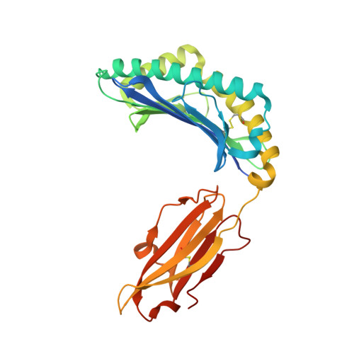
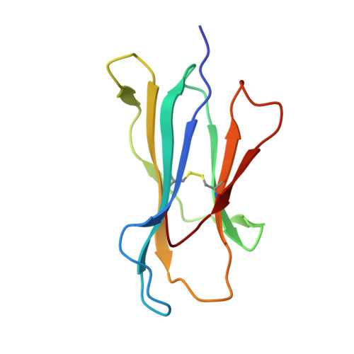
Exchange catalysis by tapasin exploits conserved and allele-specific features of MHC-I molecules.

Lan, H.Abualrous, E.T.Sticht, J.Fernandez, L.M.A.Werk, T.Weise, C.Ballaschk, M.Schmieder, P.Loll, B.Freund, C.

(2021) Nat Commun 12: 4236-4236

  • DOI: https://doi.org/10.1038/s41467-021-24401-4
  • Primary Citation Related Structures: 
    7ALO

  • PubMed Abstract: 

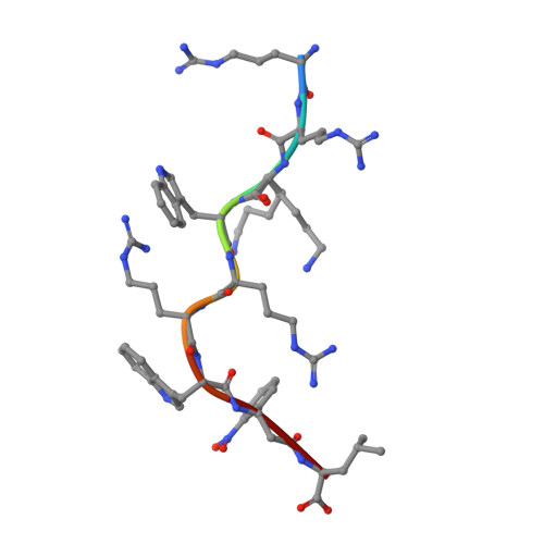
    The repertoire of peptides presented by major histocompatibility complex class I (MHC-I) molecules on the cell surface is tailored by the ER-resident peptide loading complex (PLC), which contains the exchange catalyst tapasin. Tapasin stabilizes MHC-I molecules and promotes the formation of stable peptide-MHC-I (pMHC-I) complexes that serve as T cell antigens. Exchange of suboptimal by high-affinity ligands is catalyzed by tapasin, but the underlying mechanism is still elusive. Here we analyze the tapasin-induced changes in MHC-I dynamics, and find the catalyst to exploit two essential features of MHC-I. First, tapasin recognizes a conserved allosteric site underneath the α 2-1 -helix of MHC-I, 'loosening' the MHC-I F-pocket region that accomodates the C-terminus of the peptide. Second, the scoop loop 11-20 of tapasin relies on residue L18 to target the MHC-I F-pocket, enabling peptide exchange. Meanwhile, tapasin residue K16 plays an accessory role in catalysis of MHC-I allotypes bearing an acidic F-pocket. Thus, our results provide an explanation for the observed allele-specificity of catalyzed peptide exchange.


  • Organizational Affiliation
    • Laboratory of Protein Biochemistry, Institute for Chemistry & Biochemistry, Freie Universität Berlin, Berlin, Germany.

Macromolecule Content 

  • Total Structure Weight: 96.25 kDa 
  • Atom Count: 7,344 
  • Modeled Residue Count: 768 
  • Deposited Residue Count: 802 
  • Unique protein chains: 3

Macromolecules

Find similar proteins by:|  3D Structure
Entity ID: 1
MoleculeChains  Sequence LengthOrganismDetailsImage
Lymphocyte antigen HLA-B27
A, D
292Homo sapiensMutation(s): 0 
Gene Names: HLA-B
UniProt
Find proteins for A0A2R7Z5J3 (Homo sapiens)
Explore A0A2R7Z5J3 
Go to UniProtKB:  A0A2R7Z5J3
Entity Groups
Sequence Clusters30% Identity50% Identity70% Identity90% Identity95% Identity100% Identity
UniProt GroupA0A2R7Z5J3
Sequence Annotations
Expand
Reference Sequence
Find similar proteins by:|  3D Structure
Entity ID: 2
MoleculeChains  Sequence LengthOrganismDetailsImage
Beta-2-microglobulin
B, E
100Homo sapiensMutation(s): 0 
Gene Names: B2MCDABP0092HDCMA22P
UniProt & NIH Common Fund Data Resources
Find proteins for P61769 (Homo sapiens)
Explore P61769 
Go to UniProtKB:  P61769
PHAROS:  P61769
GTEx:  ENSG00000166710 
Entity Groups
Sequence Clusters30% Identity50% Identity70% Identity90% Identity95% Identity100% Identity
UniProt GroupP61769
Sequence Annotations
Expand
Reference Sequence
Find similar proteins by:  Sequence   |   3D Structure  
Entity ID: 3
MoleculeChains  Sequence LengthOrganismDetailsImage
Vasoactive intestinal polypeptide receptor 1
C, F
9Homo sapiensMutation(s): 0 
UniProt & NIH Common Fund Data Resources
Find proteins for P32241 (Homo sapiens)
Explore P32241 
Go to UniProtKB:  P32241
PHAROS:  P32241
GTEx:  ENSG00000114812 
Entity Groups
UniProt GroupP32241
Sequence Annotations
Expand
Reference Sequence

Small Molecules

Ligands 5 Unique
IDChains Name / Formula / InChI Key2D Diagram3D Interactions
1PE

Query on 1PE


Download:Ideal Coordinates CCD File
M [auth B]PENTAETHYLENE GLYCOL
C10 H22 O6
JLFNLZLINWHATN-UHFFFAOYSA-N
PGE

Query on PGE


Download:Ideal Coordinates CCD File
N [auth B]TRIETHYLENE GLYCOL
C6 H14 O4
ZIBGPFATKBEMQZ-UHFFFAOYSA-N
TRS

Query on TRS


Download:Ideal Coordinates CCD File
L [auth B],
S [auth D]
2-AMINO-2-HYDROXYMETHYL-PROPANE-1,3-DIOL
C4 H12 N O3
LENZDBCJOHFCAS-UHFFFAOYSA-O
PEG

Query on PEG


Download:Ideal Coordinates CCD File
T [auth D]DI(HYDROXYETHYL)ETHER
C4 H10 O3
MTHSVFCYNBDYFN-UHFFFAOYSA-N
GOL

Query on GOL


Download:Ideal Coordinates CCD File
G [auth A]
H [auth A]
I [auth A]
J [auth B]
K [auth B]
G [auth A],
H [auth A],
I [auth A],
J [auth B],
K [auth B],
O [auth D],
P [auth D],
Q [auth D],
R [auth D],
U [auth E],
V [auth E],
W [auth E]
GLYCEROL
C3 H8 O3
PEDCQBHIVMGVHV-UHFFFAOYSA-N

Experimental Data & Validation

Experimental Data

  • Method: X-RAY DIFFRACTION
  • Resolution: 1.80 Å
  • R-Value Free:  0.266 (Depositor), 0.266 (DCC) 
  • R-Value Work:  0.223 (Depositor), 0.225 (DCC) 
  • R-Value Observed: 0.226 (Depositor) 
Space Group: P 1
Unit Cell:
Length ( Å )Angle ( ˚ )
a = 45.2α = 80.24
b = 69.61β = 88.362
c = 82.54γ = 89.877
Software Package:
Software NamePurpose
PHENIXrefinement
XDSdata reduction
XSCALEdata scaling
PHASERphasing

Structure Validation

View Full Validation Report


Entry History 

& Funding Information

Deposition Data


Funding OrganizationLocationGrant Number
German Research Foundation (DFG)Germany1325/17-1
German Research Foundation (DFG)GermanyTRR186 A21N

Revision History  (Full details and data files)

  • Version 1.0: 2021-06-16
    Type: Initial release
  • Version 1.1: 2021-07-21
    Changes: Data collection, Database references, Source and taxonomy, Structure summary
  • Version 2.0: 2023-11-15
    Changes: Atomic model, Data collection, Database references, Derived calculations
  • Version 2.1: 2024-01-31
    Changes: Refinement description
  • Version 2.2: 2024-10-16
    Changes: Structure summary