7AI1

Crystal structure of human MDM2-G443T RING domain homodimer bound to UbcH5B-Ub (Crystal form 2)


Experimental Data Snapshot

  • Method: X-RAY DIFFRACTION
  • Resolution: 2.07 Å
  • R-Value Free: 0.266 
  • R-Value Work: 0.212 

wwPDB Validation   3D Report Full Report


This is version 2.1 of the entry. See complete history


Literature

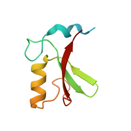
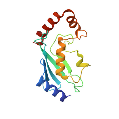
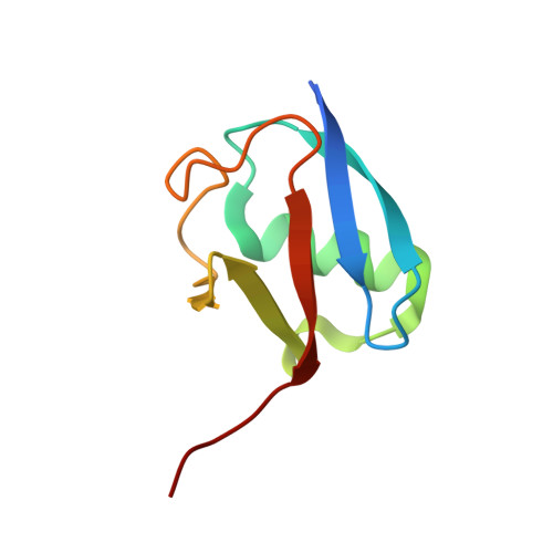
Identification of a Catalytic Active but Non-Aggregating MDM2 RING Domain Variant.

Magnussen, H.M.Huang, D.T.

(2021) J Mol Biol 433: 166807-166807

  • DOI: https://doi.org/10.1016/j.jmb.2021.166807
  • Primary Citation of Related Structures:  
    7AH2, 7AHY, 7AHZ, 7AI0, 7AI1

  • PubMed Abstract: 

    As a key regulator of the tumour suppressor protein p53, MDM2 is involved in various types of cancer and has thus been an attractive drug target. So far, small molecule design has primarily focussed on the N-terminal p53-binding domain although on-target toxicity effects have been reported. Targeting the catalytic RING domain of MDM2 resembles an alternative approach to drug MDM2 with the idea to prevent MDM2-mediated ubiquitination of p53 while retaining MDM2's ability to bind p53. The design of RING inhibitors has been limited by the extensive aggregation tendency of the RING domain, making it challenging to undertake co-crystallization attempts with potential inhibitors. Here we compare the purification profiles of the MDM2 RING domain from several species and show that the MDM2 RING domain of other species than human is much less prone to aggregate although the overall structure of the RING domain is conserved. Through sequence comparison and mutagenesis analyses, we identify a single point mutation, G443T, which greatly enhances the dimeric fraction of human MDM2 RING domain during purification. Neither does the mutation alter the structure of the RING domain, nor does it affect E2(UbcH5B)-Ub binding and activity. Hence, MDM2-G443T facilitates studies involving binding partners that would be hampered by the low solubility of the wild-type RING domain. Furthermore, it will be valuable for the development of MDM2 RING inhibitors.


  • Organizational Affiliation

    Cancer Research UK Beatson Institute, Garscube Estate, Switchback Road, Glasgow G61 1BD, United Kingdom; Institute of Cancer Sciences, University of Glasgow, Glasgow G61 1QH, United Kingdom.


Macromolecules
Find similar proteins by:  (by identity cutoff)  |  3D Structure
Entity ID: 1
MoleculeChains Sequence LengthOrganismDetailsImage
E3 ubiquitin-protein ligase Mdm2A [auth AAA],
D [auth DDD]
75Homo sapiensMutation(s): 1 
Gene Names: MDM2
EC: 2.3.2.27
UniProt & NIH Common Fund Data Resources
Find proteins for Q00987 (Homo sapiens)
Explore Q00987 
Go to UniProtKB:  Q00987
PHAROS:  Q00987
GTEx:  ENSG00000135679 
Entity Groups  
Sequence Clusters30% Identity50% Identity70% Identity90% Identity95% Identity100% Identity
UniProt GroupQ00987
Sequence Annotations
Expand
  • Reference Sequence
Find similar proteins by:  (by identity cutoff)  |  3D Structure
Entity ID: 2
MoleculeChains Sequence LengthOrganismDetailsImage
Ubiquitin-conjugating enzyme E2 D2B [auth BBB],
E [auth EEE]
147Homo sapiensMutation(s): 2 
Gene Names: UBE2D2PUBC1UBC4UBC5BUBCH4UBCH5B
EC: 2.3.2.23 (PDB Primary Data), 2.3.2.24 (PDB Primary Data)
UniProt & NIH Common Fund Data Resources
Find proteins for P62837 (Homo sapiens)
Explore P62837 
Go to UniProtKB:  P62837
PHAROS:  P62837
GTEx:  ENSG00000131508 
Entity Groups  
Sequence Clusters30% Identity50% Identity70% Identity90% Identity95% Identity100% Identity
UniProt GroupP62837
Sequence Annotations
Expand
  • Reference Sequence
Find similar proteins by:  (by identity cutoff)  |  3D Structure
Entity ID: 3
MoleculeChains Sequence LengthOrganismDetailsImage
Polyubiquitin-BC [auth CCC],
F [auth FFF]
81Homo sapiensMutation(s): 0 
Gene Names: UBB
UniProt & NIH Common Fund Data Resources
Find proteins for P0CG47 (Homo sapiens)
Explore P0CG47 
Go to UniProtKB:  P0CG47
PHAROS:  P0CG47
GTEx:  ENSG00000170315 
Entity Groups  
Sequence Clusters30% Identity50% Identity70% Identity90% Identity95% Identity100% Identity
UniProt GroupP0CG47
Sequence Annotations
Expand
  • Reference Sequence
Small Molecules
Ligands 3 Unique
IDChains Name / Formula / InChI Key2D Diagram3D Interactions
ZN
Query on ZN

Download Ideal Coordinates CCD File 
H [auth AAA],
I [auth AAA],
M [auth DDD],
N [auth DDD]
ZINC ION
Zn
PTFCDOFLOPIGGS-UHFFFAOYSA-N
EDO
Query on EDO

Download Ideal Coordinates CCD File 
J [auth BBB],
K [auth BBB]
1,2-ETHANEDIOL
C2 H6 O2
LYCAIKOWRPUZTN-UHFFFAOYSA-N
CL
Query on CL

Download Ideal Coordinates CCD File 
G [auth AAA],
L [auth DDD]
CHLORIDE ION
Cl
VEXZGXHMUGYJMC-UHFFFAOYSA-M
Experimental Data & Validation

Experimental Data

  • Method: X-RAY DIFFRACTION
  • Resolution: 2.07 Å
  • R-Value Free: 0.266 
  • R-Value Work: 0.212 
  • Space Group: P 21 21 21
Unit Cell:
Length ( Å )Angle ( ˚ )
a = 56.267α = 90
b = 80.687β = 90
c = 135.909γ = 90
Software Package:
Software NamePurpose
REFMACrefinement
xia2data reduction
xia2data scaling
PHASERphasing

Structure Validation

View Full Validation Report


Entry History & Funding Information

Deposition Data


Funding OrganizationLocationGrant Number
Cancer Research UKUnited KingdomA17196
Cancer Research UKUnited KingdomA29256
European Research Council (ERC)United Kingdom647849

Revision History  (Full details and data files)

  • Version 1.0: 2021-01-20
    Type: Initial release
  • Version 2.0: 2021-01-27
    Changes: Atomic model, Database references
  • Version 2.1: 2021-02-10
    Changes: Database references