7AEU

Pressure wave-exposed human hemoglobin: probe only data (5500 indexed images)


Experimental Data Snapshot

  • Method: X-RAY DIFFRACTION
  • Resolution: 2.54 Å
  • R-Value Free: 0.273 
  • R-Value Work: 0.207 

wwPDB Validation   3D Report Full Report


Ligand Structure Quality Assessment 


This is version 1.1 of the entry. See complete history


Literature

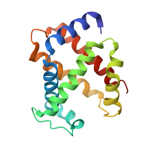
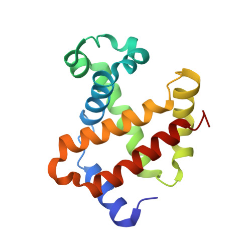
Effect of X-ray free-electron laser-induced shockwaves on haemoglobin microcrystals delivered in a liquid jet.

Grunbein, M.L.Gorel, A.Foucar, L.Carbajo, S.Colocho, W.Gilevich, S.Hartmann, E.Hilpert, M.Hunter, M.Kloos, M.Koglin, J.E.Lane, T.J.Lewandowski, J.Lutman, A.Nass, K.Nass Kovacs, G.Roome, C.M.Sheppard, J.Shoeman, R.L.Stricker, M.van Driel, T.Vetter, S.Doak, R.B.Boutet, S.Aquila, A.Decker, F.J.Barends, T.R.M.Stan, C.A.Schlichting, I.

(2021) Nat Commun 12: 1672-1672

  • DOI: https://doi.org/10.1038/s41467-021-21819-8
  • Primary Citation of Related Structures:  
    7AET, 7AEU, 7AEV

  • PubMed Abstract: 

    X-ray free-electron lasers (XFELs) enable obtaining novel insights in structural biology. The recently available MHz repetition rate XFELs allow full data sets to be collected in shorter time and can also decrease sample consumption. However, the microsecond spacing of MHz XFEL pulses raises new challenges, including possible sample damage induced by shock waves that are launched by preceding pulses in the sample-carrying jet. We explored this matter with an X-ray-pump/X-ray-probe experiment employing haemoglobin microcrystals transported via a liquid jet into the XFEL beam. Diffraction data were collected using a shock-wave-free single-pulse scheme as well as the dual-pulse pump-probe scheme. The latter, relative to the former, reveals significant degradation of crystal hit rate, diffraction resolution and data quality. Crystal structures extracted from the two data sets also differ. Since our pump-probe attributes were chosen to emulate EuXFEL operation at its 4.5 MHz maximum pulse rate, this prompts concern about such data collection.


  • Organizational Affiliation

    Max Planck Institute for Medical Research, Jahnstrasse 29, Heidelberg, Germany.


Macromolecules
Find similar proteins by:  (by identity cutoff)  |  3D Structure
Entity ID: 1
MoleculeChains Sequence LengthOrganismDetailsImage
Hemoglobin subunit alphaA [auth AAA],
C [auth CCC]
139Homo sapiensMutation(s): 0 
UniProt & NIH Common Fund Data Resources
Find proteins for P69905 (Homo sapiens)
Explore P69905 
Go to UniProtKB:  P69905
PHAROS:  P69905
Entity Groups  
Sequence Clusters30% Identity50% Identity70% Identity90% Identity95% Identity100% Identity
UniProt GroupP69905
Sequence Annotations
Expand
  • Reference Sequence
Find similar proteins by:  (by identity cutoff)  |  3D Structure
Entity ID: 2
MoleculeChains Sequence LengthOrganismDetailsImage
Hemoglobin subunit betaB [auth BBB],
D [auth DDD]
145Homo sapiensMutation(s): 0 
UniProt & NIH Common Fund Data Resources
Find proteins for P68871 (Homo sapiens)
Explore P68871 
Go to UniProtKB:  P68871
PHAROS:  P68871
GTEx:  ENSG00000244734 
Entity Groups  
Sequence Clusters30% Identity50% Identity70% Identity90% Identity95% Identity100% Identity
UniProt GroupP68871
Sequence Annotations
Expand
  • Reference Sequence
Experimental Data & Validation

Experimental Data

  • Method: X-RAY DIFFRACTION
  • Resolution: 2.54 Å
  • R-Value Free: 0.273 
  • R-Value Work: 0.207 
  • Space Group: P 21 21 21
Unit Cell:
Length ( Å )Angle ( ˚ )
a = 55.7α = 90
b = 158.1β = 90
c = 67.7γ = 90
Software Package:
Software NamePurpose
REFMACrefinement
CrystFELdata reduction
CrystFELdata scaling
REFMACphasing

Structure Validation

View Full Validation Report


Ligand Structure Quality Assessment 


Entry History 

Deposition Data

Revision History  (Full details and data files)

  • Version 1.0: 2021-02-17
    Type: Initial release
  • Version 1.1: 2021-04-07
    Changes: Database references