7A0L

Joint neutron/X-ray room temperature structure of perdeuterated Aspergillus flavus urate oxidase in complex with the 8-azaxanthine inhibitor and catalytic water bound in the peroxo hole


Experimental Data Snapshot

  • Method: X-RAY DIFFRACTION
  • Resolution: 1.33 Å
  • R-Value Free: 0.109 
  • R-Value Work: 0.102 
  • R-Value Observed: 0.102 

  • Method: NEUTRON DIFFRACTION
  • Resolution: 2.10 Å
  • R-Value Free: 0.230 
  • R-Value Work: 0.215 

wwPDB Validation   3D Report Full Report


This is version 1.1 of the entry. See complete history


Literature

Joint neutron/X-ray crystal structure of a mechanistically relevant complex of perdeuterated urate oxidase and simulations provide insight into the hydration step of catalysis.

McGregor, L.Foldes, T.Bui, S.Moulin, M.Coquelle, N.Blakeley, M.P.Rosta, E.Steiner, R.A.

(2021) IUCrJ 8: 46-59

  • DOI: https://doi.org/10.1107/S2052252520013615
  • Primary Citation of Related Structures:  
    7A0L

  • PubMed Abstract: 

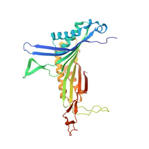
    Cofactor-independent urate oxidase (UOX) is an ∼137 kDa tetrameric enzyme essential for uric acid (UA) catabolism in many organisms. UA is first oxidized by O 2 to de-hydro-isourate (DHU) via a peroxo intermediate. DHU then undergoes hydration to 5-hy-droxy-isourate (5HIU). At different stages of the reaction both catalytic O 2 and water occupy the 'peroxo hole' above the organic substrate. Here, high-resolution neutron/X-ray crystallographic analysis at room temperature has been integrated with molecular dynamics simulations to investigate the hydration step of the reaction. The joint neutron/X-ray structure of perdeuterated Aspergillus flavus UOX in complex with its 8-azaxanthine (8AZA) inhibitor shows that the catalytic water molecule (W1) is present in the peroxo hole as neutral H 2 O, oriented at 45° with respect to the ligand. It is stabilized by Thr57 and Asn254 on different UOX protomers as well as by an O-H⋯π interaction with 8AZA. The active site Lys10-Thr57 dyad features a charged Lys10-NH 3 + side chain engaged in a strong hydrogen bond with Thr57 OG1 , while the Thr57 OG1-HG1 bond is rotationally dynamic and oriented toward the π system of the ligand, on average. Our analysis offers support for a mechanism in which W1 performs a nucleophilic attack on DHU C5 with Thr57 HG1 central to a Lys10-assisted proton-relay system. Room-temperature crystallography and simulations also reveal conformational heterogeneity for Asn254 that modulates W1 stability in the peroxo hole. This is proposed to be an active mechanism to facilitate W1/O 2 exchange during catalysis.


  • Organizational Affiliation

    Randall Centre for Cell and Molecular Biophysics, King's College London, London SE1 1UL, United Kingdom.


Macromolecules
Find similar proteins by:  (by identity cutoff)  |  3D Structure
Entity ID: 1
MoleculeChains Sequence LengthOrganismDetailsImage
Uricase302Aspergillus flavusMutation(s): 0 
Gene Names: uaZuox
EC: 1.7.3.3
UniProt
Find proteins for Q00511 (Aspergillus flavus)
Explore Q00511 
Go to UniProtKB:  Q00511
Entity Groups  
Sequence Clusters30% Identity50% Identity70% Identity90% Identity95% Identity100% Identity
UniProt GroupQ00511
Sequence Annotations
Expand
  • Reference Sequence
Small Molecules
Ligands 2 Unique
IDChains Name / Formula / InChI Key2D Diagram3D Interactions
AZA (Subject of Investigation/LOI)
Query on AZA

Download Ideal Coordinates CCD File 
B [auth A]8-AZAXANTHINE
C4 H3 N5 O2
KVGVQTOQSNJTJI-UHFFFAOYSA-N
NA
Query on NA

Download Ideal Coordinates CCD File 
C [auth A]SODIUM ION
Na
FKNQFGJONOIPTF-UHFFFAOYSA-N
Modified Residues  1 Unique
IDChains TypeFormula2D DiagramParent
CSO
Query on CSO
A
L-PEPTIDE LINKINGC3 H7 N O3 SCYS
Experimental Data & Validation

Experimental Data

  • Method: X-RAY DIFFRACTION
  • Resolution: 1.33 Å
  • R-Value Free: 0.109 
  • R-Value Work: 0.102 
  • R-Value Observed: 0.102 
  • Space Group: I 2 2 2
  • Method: NEUTRON DIFFRACTION
  • Resolution: 2.10 Å
  • R-Value Free: 0.230 
  • R-Value Work: 0.215 
  • Space Group: I 2 2 2
Unit Cell:
Length ( Å )Angle ( ˚ )
a = 80.628α = 90
b = 96.13β = 90
c = 105.397γ = 90
Software Package:
Software NamePurpose
PHENIXrefinement
LAUEGENdata processing
DIALSdata processing
SCALAdata scaling
PHENIXphasing
xia2data reduction

Structure Validation

View Full Validation Report


Entry History & Funding Information

Deposition Data


Funding OrganizationLocationGrant Number
Biotechnology and Biological Sciences Research Council (BBSRC)United KingdomBB/P000169/1

Revision History  (Full details and data files)

  • Version 1.0: 2020-12-09
    Type: Initial release
  • Version 1.1: 2021-02-10
    Changes: Database references