7A0L | pdb_00007a0l

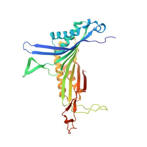
Joint neutron/X-ray room temperature structure of perdeuterated Aspergillus flavus urate oxidase in complex with the 8-azaxanthine inhibitor and catalytic water bound in the peroxo hole


Experimental Data Snapshot

  • Method: X-RAY DIFFRACTION
  • Resolution: 1.33 Å
  • R-Value Free: 
    0.109 (Depositor) 
  • R-Value Work: 
    0.102 (Depositor), 0.336 (DCC) 
  • R-Value Observed: 
    0.102 (Depositor) 

  • Method: NEUTRON DIFFRACTION
  • Resolution: 2.10 Å
  • R-Value Free: 
    0.230 (Depositor) 
  • R-Value Work: 
    0.215 (Depositor) 

Starting Model: experimental
View more details

wwPDB Validation 3D Report Full Report

Validation slider image for 7A0L

This is version 1.3 of the entry. See complete history. 

Literature

Joint neutron/X-ray crystal structure of a mechanistically relevant complex of perdeuterated urate oxidase and simulations provide insight into the hydration step of catalysis.

McGregor, L., Foldes, T., Bui, S., Moulin, M., Coquelle, N., Blakeley, M.P., Rosta, E., Steiner, R.A.

(2021) IUCrJ 8: 46-59

  • DOI: https://doi.org/10.1107/S2052252520013615
  • Primary Citation Related Structures: 
    7A0L

  • PubMed Abstract: 

    Cofactor-independent urate oxidase (UOX) is an ∼137 kDa tetrameric enzyme essential for uric acid (UA) catabolism in many organisms. UA is first oxidized by O 2 to de-hydro-isourate (DHU) via a peroxo intermediate. DHU then undergoes hydration to 5-hy-droxy-isourate (5HIU). At different stages of the reaction both catalytic O 2 and water occupy the 'peroxo hole' above the organic substrate. Here, high-resolution neutron/X-ray crystallographic analysis at room temperature has been integrated with molecular dynamics simulations to investigate the hydration step of the reaction. The joint neutron/X-ray structure of perdeuterated Aspergillus flavus UOX in complex with its 8-azaxanthine (8AZA) inhibitor shows that the catalytic water molecule (W1) is present in the peroxo hole as neutral H 2 O, oriented at 45° with respect to the ligand. It is stabilized by Thr57 and Asn254 on different UOX protomers as well as by an O-H⋯π interaction with 8AZA. The active site Lys10-Thr57 dyad features a charged Lys10-NH 3 + side chain engaged in a strong hydrogen bond with Thr57 OG1 , while the Thr57 OG1-HG1 bond is rotationally dynamic and oriented toward the π system of the ligand, on average. Our analysis offers support for a mechanism in which W1 performs a nucleophilic attack on DHU C5 with Thr57 HG1 central to a Lys10-assisted proton-relay system. Room-temperature crystallography and simulations also reveal conformational heterogeneity for Asn254 that modulates W1 stability in the peroxo hole. This is proposed to be an active mechanism to facilitate W1/O 2 exchange during catalysis.


  • Organizational Affiliation: 
    • Randall Centre for Cell and Molecular Biophysics, King's College London, London SE1 1UL, United Kingdom.

Macromolecule Content 

  • Total Structure Weight: 34.46 kDa 
  • Atom Count: 2,974 
  • Modeled Residue Count: 296 
  • Deposited Residue Count: 303 
  • Unique protein chains: 1

Macromolecules

Find similar proteins by:|  3D Structure
Entity ID: 1
MoleculeChains  Sequence LengthOrganismDetailsImage
Uricase302Aspergillus flavusMutation(s): 0 
Gene Names: uaZ, uox
EC: 1.7.3.3
UniProt
Find proteins for Q00511 (Aspergillus flavus)
Explore Q00511 
Go to UniProtKB:  Q00511
Entity Groups
Sequence Clusters30% Identity50% Identity70% Identity90% Identity95% Identity100% Identity
UniProt GroupQ00511
Sequence Annotations
Expand
Reference Sequence

Small Molecules

Ligands 2 Unique
IDChains Name / Formula / InChI Key2D Diagram3D Interactions
AZA
(Subject of Investigation/LOI)

Query on AZA


Download:Ideal Coordinates CCD File
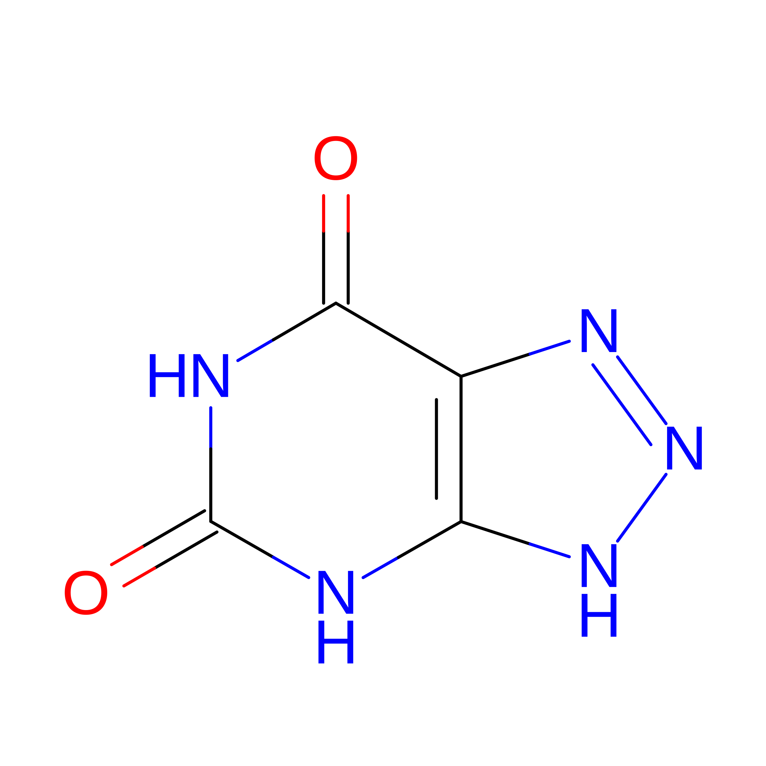
B [auth A]8-AZAXANTHINE
C4 H3 N5 O2
KVGVQTOQSNJTJI-UHFFFAOYSA-N
NA

Query on NA


Download:Ideal Coordinates CCD File
C [auth A]SODIUM ION
Na
FKNQFGJONOIPTF-UHFFFAOYSA-N
Modified Residues  1 Unique
IDChains TypeFormula2D DiagramParent
CSO
Query on CSO
A
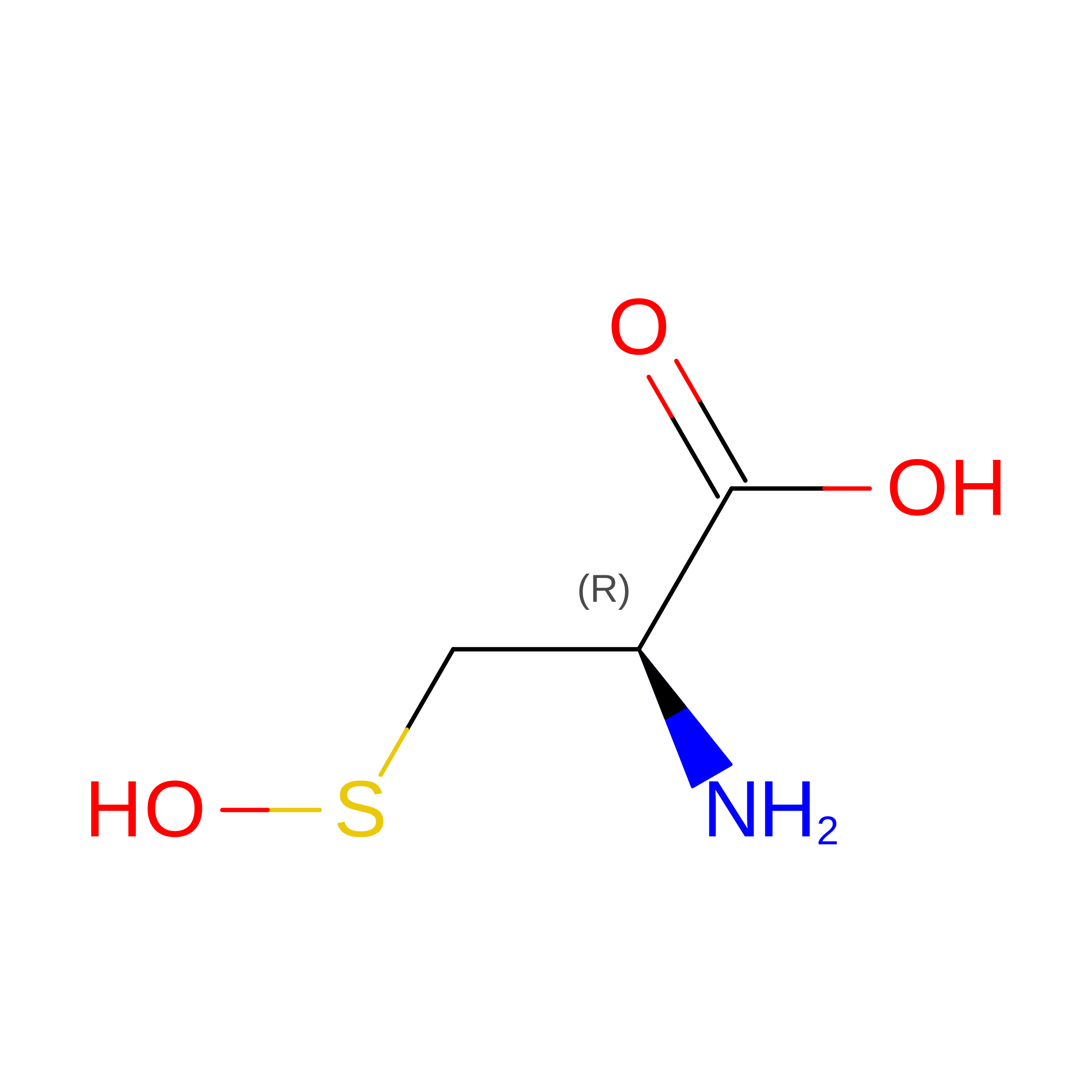
L-PEPTIDE LINKINGC3 H7 N O3 SCYS

Experimental Data & Validation

Experimental Data

  • Method: X-RAY DIFFRACTION
  • Resolution: 1.33 Å
  • R-Value Free:  0.109 (Depositor) 
  • R-Value Work:  0.102 (Depositor), 0.336 (DCC) 
  • R-Value Observed: 0.102 (Depositor) 
  • Method: NEUTRON DIFFRACTION
  • Resolution: 2.10 Å
  • R-Value Free:  0.230 (Depositor) 
  • R-Value Work:  0.215 (Depositor) 
Space Group: I 2 2 2
Unit Cell:
Length ( Å )Angle ( ˚ )
a = 80.628α = 90
b = 96.13β = 90
c = 105.397γ = 90
Software Package:
Software NamePurpose
PHENIXrefinement
LAUEGENdata processing
DIALSdata processing
SCALAdata scaling
PHENIXphasing
xia2data reduction

Structure Validation

View Full Validation Report


Entry History 

& Funding Information

Deposition Data


Funding OrganizationLocationGrant Number
Biotechnology and Biological Sciences Research Council (BBSRC)United KingdomBB/P000169/1

Revision History  (Full details and data files)

  • Version 1.0: 2020-12-09
    Type: Initial release
  • Version 1.1: 2021-02-10
    Changes: Database references
  • Version 1.2: 2024-05-01
    Changes: Data collection, Database references, Refinement description
  • Version 1.3: 2024-11-20
    Changes: Structure summary