7A0A

Crystal structure of mouse CSAD in apo form


Experimental Data Snapshot

  • Method: X-RAY DIFFRACTION
  • Resolution: 2.80 Å
  • R-Value Free: 0.292 
  • R-Value Work: 0.243 
  • R-Value Observed: 0.245 

wwPDB Validation   3D Report Full Report


This is version 1.1 of the entry. See complete history


Literature

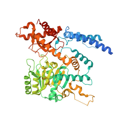
The structure of cysteine sulphinic acid decarboxylase reveals structural determinants for substrate specificity of pyridoxal phosphate-dependent decarboxylases

Mahootchi, E.Raasakka, A.Luan, W.Muruganandam, G.Loris, R.Haavik, J.Kursula, P.

To be published.

Macromolecules
Find similar proteins by:  (by identity cutoff)  |  3D Structure
Entity ID: 1
MoleculeChains Sequence LengthOrganismDetailsImage
Cysteine sulfinic acid decarboxylase
A, B, C, D
521Mus musculusMutation(s): 0 
Gene Names: Csad
EC: 4.1.1.29 (PDB Primary Data), 4.1.1.11 (PDB Primary Data)
UniProt
Find proteins for Q9DBE0 (Mus musculus)
Explore Q9DBE0 
Go to UniProtKB:  Q9DBE0
Entity Groups  
Sequence Clusters30% Identity50% Identity70% Identity90% Identity95% Identity100% Identity
UniProt GroupQ9DBE0
Sequence Annotations
Expand
  • Reference Sequence
Experimental Data & Validation

Experimental Data

  • Method: X-RAY DIFFRACTION
  • Resolution: 2.80 Å
  • R-Value Free: 0.292 
  • R-Value Work: 0.243 
  • R-Value Observed: 0.245 
  • Space Group: P 1 21 1
Unit Cell:
Length ( Å )Angle ( ˚ )
a = 73.06α = 90
b = 114.88β = 95.804
c = 113.76γ = 90
Software Package:
Software NamePurpose
PHENIXrefinement
XDSdata processing
XSCALEdata scaling
PHASERphasing

Structure Validation

View Full Validation Report


Entry History 

Deposition Data

Revision History  (Full details and data files)

  • Version 1.0: 2021-08-18
    Type: Initial release
  • Version 1.1: 2024-01-31
    Changes: Data collection, Refinement description